Review
. 2022 Aug 17;14(16):3965.
doi: 10.3390/cancers14163965.

The Role of CT in the Staging and Follow-Up of Testicular Tumors: Baseline, Recurrence and Pitfalls

Affiliations
Review

The Role of CT in the Staging and Follow-Up of Testicular Tumors: Baseline, Recurrence and Pitfalls

Thibaut Pierre et al. Cancers (Basel). .

Abstract

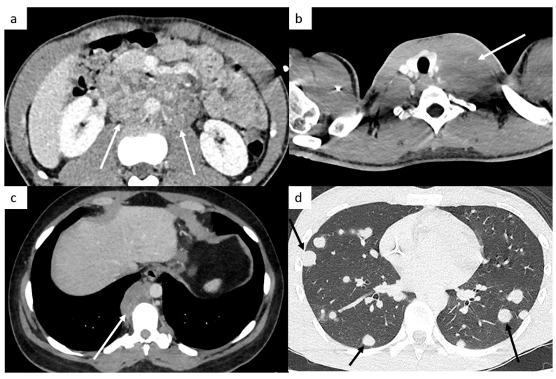
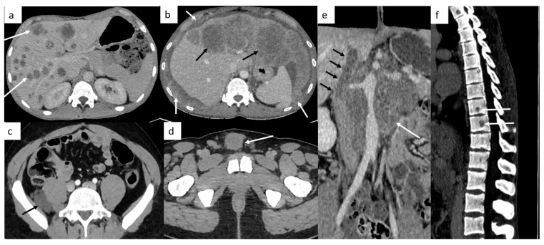
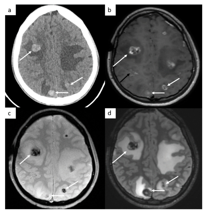
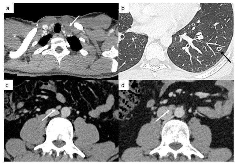
Ultrasound imaging of the testis represents the standard-of-care initial imaging for the diagnosis of TGCT, whereas computed tomography (CT) plays an integral role in the initial accurate disease staging (organ-confined, regional lymph nodes, or sites of distant metastases), in monitoring the response to therapy in patients who initially present with non-confined disease, in planning surgical approaches for residual masses, in conducting follow-up surveillance and in determining the extent of recurrence in patients who relapse after treatment completion. CT imaging has also an important place in diagnosing complications of treatments. The aims of this article are to review these different roles of CT in primary TGCT and focus on different pitfalls that radiologists need to be aware of.

Keywords: computed tomography (CT); germ cells tumors (GCT); oncologic diagnostic imaging; retroperitoneal masses; seminomatous and nonseminomatous GCT; testicular cancer (TC).


Conflict of interest statement

Karim Fizazi: Participation on advisory boards and talks for: Amgen, Astellas, Astrazeneca, Bayer, Clovis, Daiichi Sankyo, Janssen, MSD, Novartis/AAA, Pfizer, Sanofi. Honoraria go to Gustave Roussy and my institution. Participation on advisory boards with personal honorarium for CureVac and Orion. Other authors declare no conflict of interest.

Figures

Figure 1
Figure 1
Most common sites of lymph nodes and metastases in testicular cancer. Abdominal computed tomography (CT) shows retroperitoneal paraaortic lymph nodes (a, arrow). Thoracic CT reveals left supraclavicular lymph node (b, arrow). Thoracic CT demonstrates posterior mediastinal lymph nodes (c). Thoracic CT scan shows lung metastases appearing as multiple lung nodules (d).
Figure 2
Figure 2
Other sites of metastatic disease. In advanced stages, abdominal CT can show different sites of metastases including liver ((a,b), black arrows), peritoneal carcinomatosis ((b), white arrows), retroperitoneal carcinomatosis (c), cutaneous metastases in front of pubis (d). In case of large retroperitoneal lymph nodes ((e), white arrows), a tumor thrombus in the inferior vena cava can be found ((e), black arrows). Spinal bone CT shows several vertebral metastases (f).
Figure 3
Figure 3
Brain metastases. Cerebral CT brain demonstrates several hemorrhagic metastases (a). MRI confirms the presence of several metastases with peripheral enhancement ((b), white arrows). They are hypointense on SWI-weighted images (c), with surrounding edema (d) on FLAIR-weighted images. In this case, a leptomeningitis is associated ((b), black arrow).
Figure 4
Figure 4
Small disease. Thoracic CT reveals a small left supraclavicular lymph node ((a), arrow). Lung metastases can appear as different features such as small nodules which may be excavated (b). Abdominal CT demonstrates a small interaorticocaval lymph node (c) and the repetition of the CT 6 months later shows an increasing size of this lymph node, a finding indicative of involvement (d).
Figure 5
Figure 5
Post-chemotherapy lymph node changes. Abdominal CT reveals a left paraaortic lymph node measuring 27 mm (short axis) at initial staging ((a), arrow). After chemotherapy, CT shows a decrease in the size of this lymph node measuring 14 mm (short axis) ((b), arrow). Lymphadenectomy was performed, and histological examination of the surgical retroperitoneal resection specimen revealed teratomatous tissue and inflammatory changes, without any residual nodal invasion. Imaging in patient with left testicle seminoma (c,d). Abdominal CT demonstrates extensive retroperitoneal lymph nodes encasing the aorta at initial staging (c); follow-up evaluation after chemotherapy shows significant treatment response: lymph nodes decrease in size (<3 cm) and the FDG-PET CT confirms an excellent response, without increased activity in residual mass (d).
Figure 6
Figure 6
Teratoma and growing teratoma. Imaging in patient with a mature teratoma of the left testicle. Abdominal CT reveals a retroperitoneal mass ((a,b) white arrows) with fat tissue ((a,b) black arrows) corresponding to a teratoma. Abdominal CT shows a left paraaortic lymph node (c) with an increasing size during chemotherapy with classical features of growing teratoma: better circumscribed margins, expanding cystic and necrotic appearance of the lesion (d).
Figure 7
Figure 7
False-positive diagnoses. Thoracic CT shows a low-density mass below the carina in the middle mediastinum (a). A metastatic lymph node was suspected; however, this patient did not have any retroperitoneal lymph nodes. An MRI shows a well-circumscribed mass below the carina that appears hyperintense on T1-weighted images, hyperintense on T2-weighted images (b), without hypersignal on diffusion-weighted images. The FDG-PET CT confirms the absence of increased activity in this mass (c). It is a bronchogenic cyst. Post-surgical abdominal CT demonstrates a left paraaortic lymphocele (d) decreasing in size 3 months later, (e) but two years later, a low-density retrocaval mass appeared corresponding to teratoma (f). It shows that it can be difficult to differentiate teratoma from lymphocele, especially without prior CT to assess evolutions.
Figure 8
Figure 8
Post-treatment complications. Thoracic CT shows a bleomycin-induced interstitial pneumonitis (a) affecting left upper and lower lobes. Postoperative abdominal CT shows chylous ascites (b).

References

    1. Garner M.J., Turner M.C., Ghadirian P., Krewski D. Epidemiology of testicular cancer: An overview. Int. J. Cancer. 2005;116:331–339. doi: 10.1002/ijc.21032. - DOI - PubMed
    1. Siegel R.L., Miller K.D., Fuchs H.E., Jemal A. Cancer Statistics, 2021. CA Cancer J. Clin. 2021;71:7–33. doi: 10.3322/caac.21654. - DOI - PubMed
    1. World Cancer Research Fund Worldwide Cancer Data: Global Cancer Statistics for the Most Common Cancers. [(accessed on 21 January 2022)]. Available online: https://www.wcrf.org/dietandcancer/cancer-trends/worldwide-cancer-data.
    1. Jacobsen G.K., Barlebo H., Olsen J., Schultz H.P., Starklint H., Søgaard H., Væth M., Group D.S. Testicular Germ Cell Tumours in Denmark 1976–1980 Pathology of 1058 Consecutive Cases. Acta Radiol. Oncol. 1984;23:239–247. doi: 10.3109/02841868409136019. - DOI - PubMed
    1. Rajpert-De Meyts E., McGlynn K.A., Okamoto K., Jewett M.A., Bokemeyer C. Testicular germ cell tumours. Lancet. 2016;387:1762–1774. doi: 10.1016/S0140-6736(15)00991-5. - DOI - PubMed

LinkOut - more resources