Review
. 2022 Aug 20;14(16):4020.
doi: 10.3390/cancers14164020.

Tumour Derived Extracellular Vesicles: Challenging Target to Blunt Tumour Immune Evasion

Affiliations
Review

Tumour Derived Extracellular Vesicles: Challenging Target to Blunt Tumour Immune Evasion

Tatiana Lopatina et al. Cancers (Basel). .

Abstract

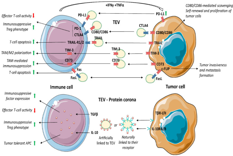
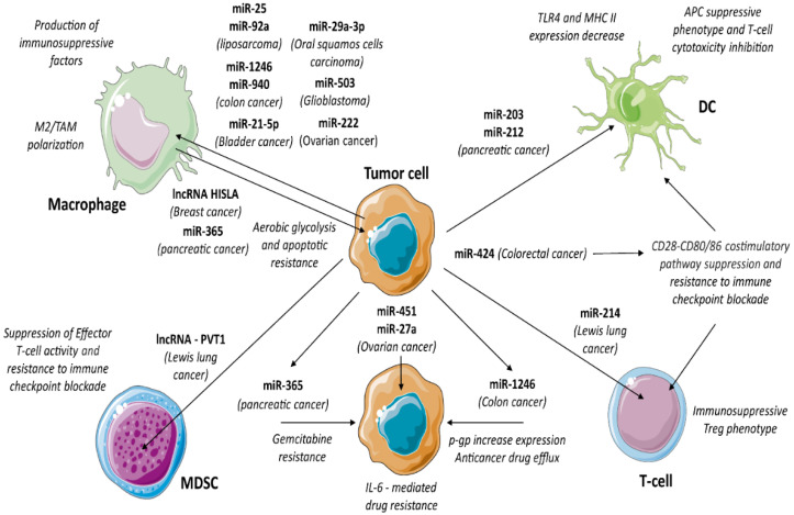
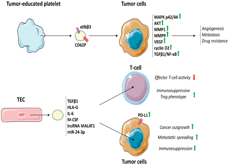
Control of the immune response is crucial for tumour onset and progression. Tumour cells handle the immune reaction by means of secreted factors and extracellular vesicles (EV). Tumour-derived extracellular vesicles (TEV) play key roles in immune reprogramming by delivering their cargo to different immune cells. Tumour-surrounding tissues also contribute to tumour immune editing and evasion, tumour progression, and drug resistance via locally released TEV. Moreover, the increase in circulating TEV has suggested their underpinning role in tumour dissemination. This review brings together data referring to TEV-driven immune regulation and antitumour immune suppression. Attention was also dedicated to TEV-mediated drug resistance.

Keywords: cell-to-cell communication; exosomes; extracellular vesicles; tumour antigens; tumour immune editing; tumour immune suppression.


Conflict of interest statement

The authors declare no conflict of interest.

Figures

Figure 1
Figure 1
TEV markers relevant for immune regulation. TEV from cancer cells regulate the immune system by acting on different cell types. In particular, TEV express a large subset of molecules able to trigger tumour immune evasion. Indeed, the interaction between ICAM-1, embedded within tetraspanin domains (CD63, CD81 and CD9) in TEV and LFA1 in leucocytes, is essential for TEV uptake by leucocytes. Moreover, the cross talk between TEV DAMPs/PAMPs and dsDNA/RNA molecules and TLR in tumour cells stimulates the expression of genes involved in immune suppression. However, TLR can be also carried by TEV initiating signalling that impairs the antitumour immune response. Furthermore, HSP70 in TEV interacts with TLR2 on MDSC, boosting p-STAT3 expression and IL-6 production, while HSP27/60 interacting with TLR in TAM promotes differentiation of pro-tumoural M2 and the production of immune suppressive factors. α5β1 and αvβ3 integrins in TEV interact with fibronectin on fibroblasts and mediate signalling in recipient cells, supporting organotrophic metastatic spread. Licensed under a Creative Commons Attribution 3.0 Unported License. https://smart.servier.com (access on 15 August 2022).
Figure 2
Figure 2
TEV as immunosuppressive shuttles. TEV from tumour cells carry various immune checkpoint molecules, which impair the antitumour immune response. In particular, activated T-cell producing IFNγ and TNFα enhance PD-L1 expression in tumour cells, which in turn secrete TEV expressing PD-L1, which interacting with PD-1, interferes with effector T-cell activity and drives Treg expansion. As PD-1, CTLA4 after binding to TEV expressing CD80/CD86 suppresses the immune response and T-cell-mediated cytotoxic activity. On the other hand, TEV carrying CTLA4 boost CD80/CD86 scavenging and induce proliferation of targeted tumour cells. Moreover, TIM-3+ TEV are uptaken by TAM and induce M2 polarisation. Similarly, CD73+ TEV upregulate the expression of immunosuppressive factors in TAM by activating the NF-κB pathway. Furthermore, FasL and TRAIL TEV content has immunosuppressive factors, by binding receptors Fas and TRAIL-R1/2, respectively, to immune cells, thereby promoting target cell apoptosis. In addition, FasL interaction with c-FLIP expressed by tumour cells promotes tumour invasiveness and metastasis formation. The so-called “Protein corona” describes the presence of TEV membrane proteins artificially linked to the membrane of TEV or naturally linked to their receptors. IL-10 and TGFβ expressed by TEV boost the immune suppression. https://smart.servier.com (access on 15 August 2022).
Figure 3
Figure 3
TEV non-coding RNA content. TEV contain and deliver a large subset of microRNAs and lncRNAs involved in immune suppression. In particular, specific miRNAs act on specific cell type. Indeed, miR-1246 and miR-940 (colon cancer), miR-222 (ovarian cancer), miR-29a-3p (oral squamous cell carcinoma), miR-21-5p (bladder cancer), miR-503 (glioblastoma), miR-25 and miR-92a (liposarcoma) stimulate macrophages to shift towards M2 phenotype. On the other hand, TEV derived from M2 macrophage carrying miR-365 (pancreatic cancer) and lncRNA HISLA (breast cancer) promote aerobic glycolysis and resistance to apoptosis in tumour cells. miR-424 (colorectal cancer) induces resistance to immune checkpoint blockade in tumour infiltrating DC and T-cells. Interestingly, miR-203 (pancreatic cancer) inhibits the expression of TLR4, while miR-212 modulates the expression of MHC II in recipient DC, which in turn induces a suppressive phenotype and inhibits T-cell cytotoxic activity. miR-214 (Lewis lung cancer) promotes the shift of T-cells towards Treg supporting immune suppression. Moreover, lncRNA-PVT1 (Lewis lung cancer) contributes to suppression CTL and resistance to immune checkpoint blockade. However, miRNAs also play a crucial role in drug resistance. Thus, miR-451 and miR-27a (ovarian cancer) enhance IL-6-mediated drug resistance, miR-365 (pancreatic cancer) induces gemcitabine resistance, while miR-1246 (colorectal cancer) upregulates p-gp to boost drug efflux. https://smart.servier.com (access on 15 August 2022).
Figure 4
Figure 4
TEV released by tumour-educated platelets and tumour endothelial cells. Tumour-educated platelets produce TEV, expressing P-selectin (CD62P) and integrin αIIbβ3, stimulate MAPK p42/44 and AKT, and increase the expression of MMP1, MMP9, VEGF and cyclin D2. Moreover, TEV induce EMT by activating TGFβ1 and NF-κB signalling in tumour cells. All these signals increase angiogenesis, metastasis formation and drug resistance. On the other hand, TEC-derived TEV contain pro-oncogenic proteins and RNAs involved in immune regulation (TGFβ1, HLA-G, IL-6, M-CSF, as well as lncRNA MALAT1, and miR-24-3p), which, by increasing PD-L1 in tumour cells, boost cancer outgrowth, metastatic spread and immunosuppression. https://smart.servier.com (access on 15 August 2022).

Similar articles

Cited by

References

    1. Whiteside T.L. Exosomes and tumor-mediated immune suppression. J. Clin. Invest. 2016;126:1216–1223. doi: 10.1172/JCI81136. - DOI - PMC - PubMed
    1. Hoshino A., Kim H.S., Bojmar L., Gyan K.E., Cioffi M., Hernandez J., Zambirinis C.P., Rodrigues G., Molina H., Heissel S., et al. Extracellular Vesicle and Particle Biomarkers Define Multiple Human Cancers. Cell. 2020;182:1044–1061.e18. doi: 10.1016/j.cell.2020.07.009. - DOI - PMC - PubMed
    1. Slingluff C.L., Jr., Cox A.L., Stover J.M., Jr., Moore M.M., Hunt D.F., Engelhard V.H. Cytotoxic T-Lymphocyte Response to Autologous Human Squamous Cell Cancer of the Lung: Epitope Reconstitution with Peptides Extracted from HLA-Aw681. Cancer Res. 1994;54:2731–2737. - PubMed
    1. Wolfers J., Lozier A., Raposo G., Regnault A., Théry C., Masurier C., Flament C., Pouzieux S., Faure F., Tursz T., et al. Tumor-derived exosomes are a source of shared tumor rejection antigens for CTL cross-priming. Nat. Med. 2001;7:297–303. doi: 10.1038/85438. - DOI - PubMed
    1. Echchakir H., Vergnon I., Dorothée G., Grunenwald D., Chouaib S., Mami-Chouaib F. Evidence for in situ expansion of diverse antitumor-specific cytotoxic T lymphocyte clones in a human large cell carcinoma of the lung. Int. Immunol. 2000;12:537–546. doi: 10.1093/intimm/12.4.537. - DOI - PubMed

Grants and funding

LinkOut - more resources