

ADC Values of Cytologically Benign and Cytologically Malignant 18 F-FDG PET-Positive Lymph Nodes of Head and Neck Squamous Cell Carcinoma
- PMID: 36011013
- PMCID: PMC9406365
- DOI: 10.3390/cancers14164019
ADC Values of Cytologically Benign and Cytologically Malignant 18 F-FDG PET-Positive Lymph Nodes of Head and Neck Squamous Cell Carcinoma
Abstract
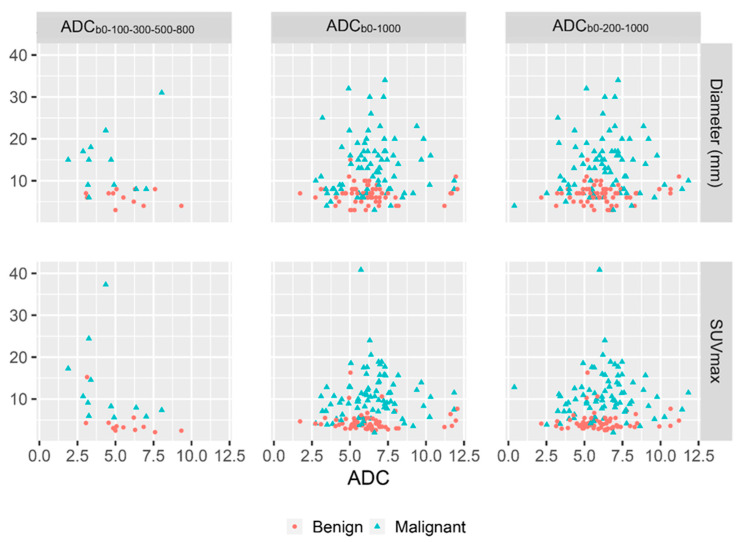
Nodal staging (N-staging) in head and neck squamous cell carcinoma (HNSCC) is essential for treatment planning and prognosis. 18F-fluordeoxyglucose positron emission tomography (FDG-PET) has high performance for N-staging, although the distinction between cytologically malignant and reactive PET-positive nodes, and consequently, the selection of nodes for ultrasound-guided fine needle aspiration cytology (USgFNAC), is challenging. Diffusion-weighted magnetic resonance imaging (DW-MRI) can help to detect nodal metastases. We aim to investigate the potential of the apparent diffusion coefficient (ADC) as a metric to distinguish between cytologically reactive and malignant PET-positive nodes in order to improve node selection criteria for USgFNAC. PET-CT, real-time image-fused USgFNAC and DW-MRI to calculate ADC were available for 78 patients offered for routine N-staging. For 167 FDG-positive nodes, differences in the ADC between cytologically benign and malignant PET-positive nodes were evaluated, and both were compared to the ADC values of PET-negative reference nodes. Analyses were also performed in subsets of nodes regarding HPV status. A mild negative correlation between SUVmax and ADC was found. No significant differences in ADC values were observed between cytologically malignant and benign PET-positive nodes overall. Within the subset of non-HPV-related nodes, ADCb0-200-1000 was significantly lower in cytologically malignant PET-positive nodes when compared to benign PET-positive nodes. ADCb0-1000 and ADCb0-200-1000 were significantly lower (p = 0.018, 0.016, resp.) in PET-negative reference nodes than in PET-positive nodes. ADC was significantly higher in PET-negative reference nodes than in PET-positive nodes. The non-HPV-related subgroup showed significantly (p = 0.03) lower ADC values in cytologically malignant than in cytologically benign PET-positive nodes, which should help inform the node selection procedure for puncture.
Keywords: DW-MRI; FDG-PET; head and neck oncology; lymph node; real-time image fusion.
Conflict of interest statement
The authors have no conflict of interest to declare that is relevant to the content of this article.
Figures
Similar articles
-
SUVmax values at FDG PET-CT to predict malignancy in lymph nodes aspirated by real time image fused USgFNAC in head and neck squamous cell carcinoma.Am J Nucl Med Mol Imaging. 2021 Jun 15;11(3):178-187. eCollection 2021. Am J Nucl Med Mol Imaging. 2021. PMID: 34234996 Free PMC article.
-
Prediction of ultrasound guided fine needle aspiration cytology results by FDG PET-CT for lymph node metastases in head and neck squamous cell carcinoma patients.Acta Oncol. 2018 Dec;57(12):1687-1692. doi: 10.1080/0284186X.2018.1529426. Epub 2018 Nov 29. Acta Oncol. 2018. PMID: 30489180
-
Correlation of the apparent diffusion coefficient (ADC) with the standardized uptake value (SUV) in lymph node metastases of non-small cell lung cancer (NSCLC) patients using hybrid 18F-FDG PET/MRI.PLoS One. 2015 Jan 9;10(1):e0116277. doi: 10.1371/journal.pone.0116277. eCollection 2015. PLoS One. 2015. PMID: 25574968 Free PMC article.
-
Real-Time Ultrasound Image Fusion with FDG-PET/CT to Perform Fused Image-Guided Fine-Needle Aspiration in Neck Nodes: Feasibility and Diagnostic Value.AJNR Am J Neuroradiol. 2021 Mar;42(3):566-572. doi: 10.3174/ajnr.A6938. Epub 2021 Jan 28. AJNR Am J Neuroradiol. 2021. PMID: 33509917 Free PMC article.
-
Positron emission tomography using [18F]fluorodeoxyglucose (FDG-PET) in the clinically negative neck: is it likely to be superior?Eur Arch Otorhinolaryngol. 2004 Oct;261(9):479-83. doi: 10.1007/s00405-003-0727-3. Epub 2003 Dec 17. Eur Arch Otorhinolaryngol. 2004. PMID: 14685887
References
-
- Singh P., Contente M., Bennett B., Hall J., Bailey H., Bailey A., Zarrelli L., Sanchez C.P. Real-World Treatment Patterns and Outcomes in Patients with Head and Neck Cancer: Point-in-Time Survey of Oncologists in Italy and Spain. Adv. Ther. 2021;38:4722–4735. doi: 10.1007/s12325-021-01851-2. - DOI - PubMed
LinkOut - more resources
Full Text Sources
