. 2022 Aug 9;23(16):8854.
doi: 10.3390/ijms23168854.

New Pyrrole Derivatives as Promising Biological Agents: Design, Synthesis, Characterization, In Silico, and Cytotoxicity Evaluation

Affiliations

New Pyrrole Derivatives as Promising Biological Agents: Design, Synthesis, Characterization, In Silico, and Cytotoxicity Evaluation

Beatrice-Cristina Ivan et al. Int J Mol Sci. .

Abstract

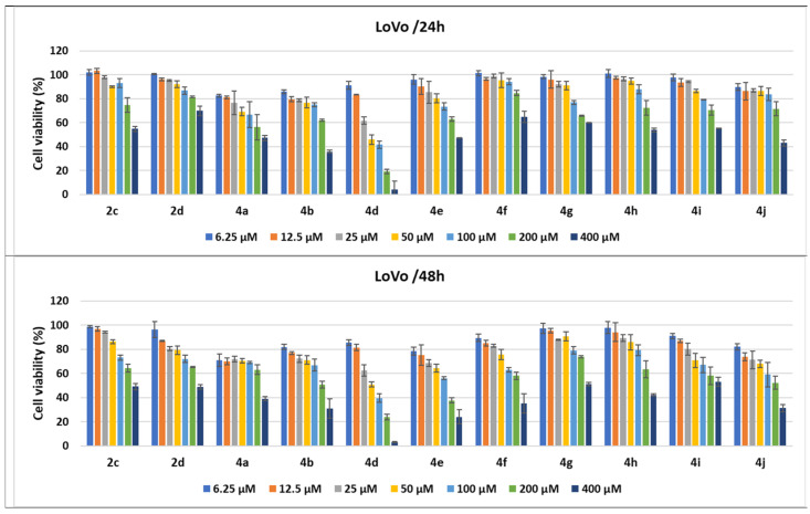
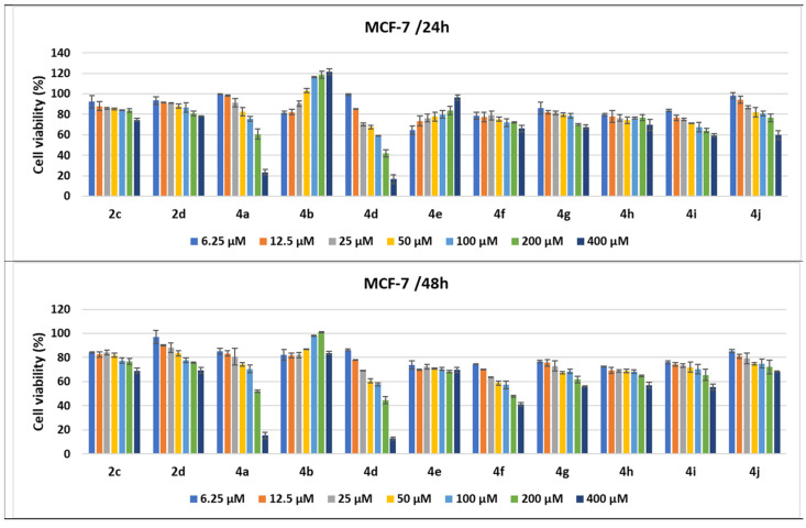
The current study describes the synthesis, physicochemical characterization and cytotoxicity evaluation of a new series of pyrrole derivatives in order to identify new bioactive molecules. The new pyrroles were obtained by reaction of benzimidazolium bromide derivatives with asymmetrical acetylenes in 1,2-epoxybutane under reflux through the Huisgen [3 + 2] cycloaddition of several ylide intermediates to the corresponding dipolarophiles. The intermediates salts were obtained from corresponding benzimidazole with bromoacetonitrile. The structures of the newly synthesized compounds were confirmed by elemental analysis, spectral techniques (i.e., IR, 1H-NMR and 13C-NMR) and single-crystal X-ray analysis. The cytotoxicity of the synthesized compounds was evaluated on plant cells (i.e., Triticum aestivum L.) and animal cells using aquatic crustaceans (i.e., Artemia franciscana Kellogg and Daphnia magna Straus). The potential antitumor activity of several of the pyrrole derivatives was studied by performing in vitro cytotoxicity assays on human adenocarcinoma-derived cell lines (i.e., LoVo (colon), MCF-7 (breast), and SK-OV-3 (ovary)) and normal human umbilical vein endothelial cells (HUVECs). The obtained results of the cytotoxicity assessment indicated that the tested compounds had nontoxic activity on Triticum aestivum L., while on Artemia franciscana Kellogg nauplii, only compounds 2c and 4c had moderate toxicity. On Daphnia magna, 4b and 4c showed high toxicity; 2a, 2b, and 2c moderate to high toxicity; only 4a and 4d were nontoxic. The compound-mediated cytotoxicity assays showed that several pyrrole compounds demonstrated dose- and time-dependent cytotoxic activity against all tested tumor cell lines, the highest antitumor properties being achieved by 4a and its homologue 4d, especially against LoVo colon cells.

Keywords: X-ray diffraction; [3 + 2] cycloaddition; antitumor activity; cytotoxicity; dipolarophile alkynes; pyrroles.


Conflict of interest statement

The authors declare no conflict of interest.

Figures

Figure 1
Figure 1
Structure of anticancer drugs sharing the pyrrole ring.
Scheme 1
Scheme 1
The synthesis of the new pyrroles, 4ad, from benzimidazole derivatives.
Scheme 2
Scheme 2
The probable mechanism for the formation of the new pyrroles 4ad.
Figure 2
Figure 2
X-ray molecular structure of compound 4d with atom labeling and thermal ellipsoids at the 40% level. Intramolecular H-bond parameters: C2-H···N1 [C2-H 0.93 Å, H···N1 2.73 Å, C2···N1 3.608 Å, ∠ C2HN1 158.3°].
Figure 3
Figure 3
A view of a 1D supramolecular chain in the crystal structure of 4d (for A molecules). Nonrelevant H atoms were omitted. H-bonds parameters: N3-H···O1 [N3-H 0.86 Å, H···O1 2.15 Å, N3···O1(1 − x, −y, 1 − z) 2.873(3) Å, ∠ N3HO1 141.3°]; C18-H···N1 [C18-H 0.93 Å, H···N1 2.59 Å, C18···N1(−x, −y, 1 − z) 3.384(4) Å, ∠ N3HO1 144.0°].
Figure 4
Figure 4
Interaction plot showing the interaction effects between the tested compounds and the concentration for the Triticum phytotoxicity test.
Figure 5
Figure 5
Violin plots showing the variations in Triticum’s main rootlet length under the influence of the substances tested at different concentration levels and day of measurement.
Figure 6
Figure 6
(a) Nuclei with hypertrophied nucleoli observed in rootlets treated with compound 2c (concentration of 1000 μM); (b) metaphase in tropokinesis and normal telophases observed in rootlets treated with compound 4c (concentration of 1000 μM); (c) telophase with chromosomal bridges observed in rootlets treated with compound 4a (concentration of 10 μM); (d) telophase with delayed chromosomes, metaphases, and interphases observed in rootlets treated with compound 2b (concentration of 100 μM). All microphotographs were taken with a lens magnified by 40×.
Figure 7
Figure 7
The lethality curves for the replicates and global models for compounds 2c and 4c.
Figure 8
Figure 8
Daphnia magna lethality curves for the tested compounds: (a) 2a; (b) 2b; (c) 2c; (d) 4a; (e) 4b; (f) 4c; (g) 4d; (h) indomethacin; error bars represent the standard error of the mean.
Figure 9
Figure 9
The structure of the pyrrole derivatives tested for antitumor activity.
Figure 10
Figure 10
Antitumor effect of pyrrole derivatives against LoVo colon cancer cells. The viability of the LoVo cells was measured after treatments with scalar concentrations of pyrrole compounds for 24 or 48 h and compared to that of untreated control cells. Data are expressed as the mean values ± standard deviations (SDs) of three different experiments (n = 3).
Figure 11
Figure 11
Antitumor effect of pyrrole derivatives against MCF-7 breast cancer cells. The viability of the MCF-7 cells was measured after treatments with scalar concentrations of pyrrole compounds for 24 or 48 h and compared to that of untreated control cells. Data are expressed as the mean value ± standard deviations (SDs) of three different experiments (n = 3).
Figure 12
Figure 12
Antitumor effect of pyrrole derivatives against SK-OV-3 ovary cancer cells. The viability of the SK-OV-3 cells was measured after treatments with scalar concentrations of pyrrole compounds for 24 or 48 h and compared to that of untreated control cells. Data are expressed as the mean values ± standard deviations (SDs) of three different experiments (n = 3).
Figure 13
Figure 13
Antitumor effect of tested compounds against HUVECs. The cell viability was measured after HUVEC treatments with scalar concentrations of the tested compounds for 24 or 48 h and compared to that of untreated control cancer cells. Data are expressed as the mean values ± standard deviations (SDs) of three different experiments (n = 3).
Figure 14
Figure 14
Antitumor effect of oncolitical control drugs against cancer vs. normal cell lines. The cell viability was measured after cell treatments with scalar concentrations of drugs for 24 or 48 h and compared to that of untreated control cells. Data are expressed as the mean values ± standard deviations (SDs) of three different experiments (n = 3).

References

    1. Sung H., Ferlay J., Siegel R.L., Laversanne M., Soerjomataram I., Jemal A., Bray F. Global Cancer Statistics 2020: GLOBOCAN Estimates of Incidence and Mortality Worldwide for 36 Cancers in 185 Countries. CA Cancer J. Clin. 2021;71:209–249. doi: 10.3322/caac.21660. - DOI - PubMed
    1. Mihaila M., Hotnog C.M., Bostan M., Munteanu A.C., Vacaroiu I.A., Brasoveanu L.I., Uivarosi V. Anticancer activity of some ruthenium (III) complexes with quinolone antibiotics: In vitro cytotoxicity, cell cycle modulation, and apoptosis-inducing properties in LoVo colon cancer cell line. Appl. Sci. 2021;11:8594. doi: 10.3390/app11188594. - DOI
    1. Tomi I.H.R., Tomma J.H., Al-Daraji A.H.R., Al-Dujaili A.H. Synthesis, characterization and comparative study the microbial activity of some heterocyclic compounds containing oxazole and benzothiazole moieties. J. Saudi Chem. Soc. 2015;19:392–398. doi: 10.1016/j.jscs.2012.04.010. - DOI
    1. Li Petri G., Spanò V., Spatola R., Holl R., Raimondi M.V., Barraja P., Montalbano A. Bioactive pyrrole-based compounds with target selectivity. Eur. J. Med. Chem. 2020;208:112783. doi: 10.1016/j.ejmech.2020.112783. - DOI - PMC - PubMed
    1. Vitaku E., Smith D.T., Njardarson J.T. Analysis of the structural diversity, substitution patterns, and frequency of nitrogen heterocycles among U.S. FDA approved pharmaceuticals. J. Med. Chem. 2014;57:10257–10274. doi: 10.1021/jm501100b. - DOI - PubMed

LinkOut - more resources