Case Reports
. 2022 Aug 10;23(16):8882.
doi: 10.3390/ijms23168882.

Analysis of the Chemical Composition and Morphological Characterization of Tissue Osseointegrated to a Dental Implant after 5 Years of Function

Affiliations
Case Reports

Analysis of the Chemical Composition and Morphological Characterization of Tissue Osseointegrated to a Dental Implant after 5 Years of Function

Josefa Alarcón Apablaza et al. Int J Mol Sci. .

Abstract

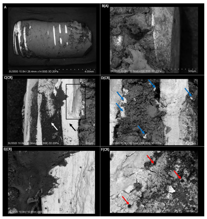
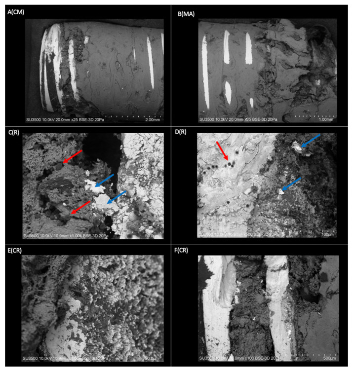
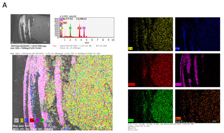
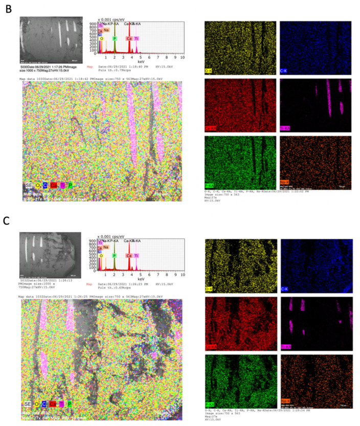
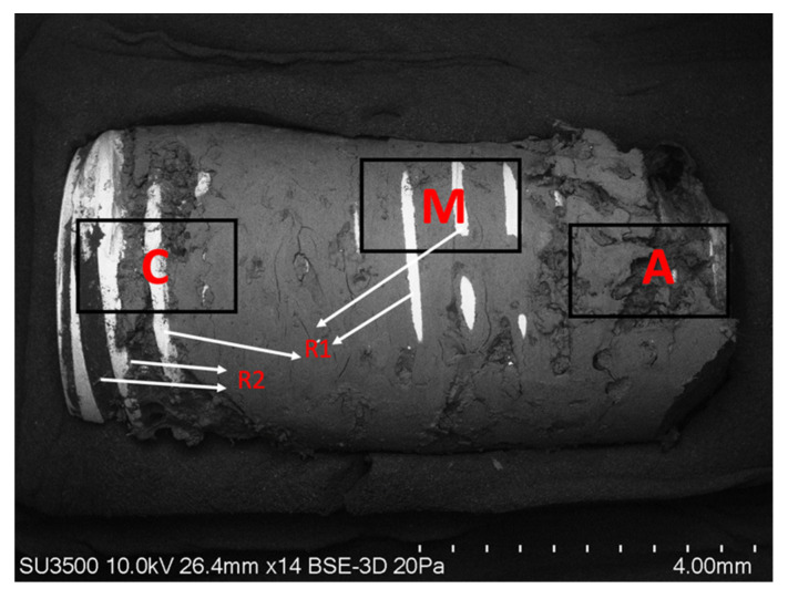
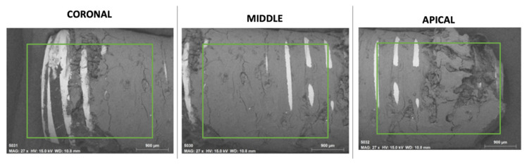
Osseointegration implies the coexistence of a biocompatible implant subjected to masticatory loads and living bone tissue adhered to its surface; this interaction is a critical process for the success of implants. The objective of this work is to analyze the osseoformation and osseointegration of a dental implant in operation for 5 years microscopically through morphological analysis of the surface and chemical composition through a variable pressure scanning electron microscope (VP-SEM) and energy dispersive X-ray spectrometry (EDX). The chemical composition and general characteristics of the structural morphology of random areas of the surfaces of an osseointegrated dental implant from an ex vivo sample were analyzed. On the surface of the implant free of bone tissue, titanium (TI) was mainly identified in the area of the implant threads and carbon (C) in the depth of the implant threads. Phosphorus (P), calcium (Ca), oxygen (O), carbon (C), with dense and homogeneous distribution, and, to a lesser extent, sodium (Na) were detected on the bone surface around the contour of the implant. Regarding the morphological characteristics of the implant surface, a rough structure with some irregularities and detachments of the implant lodged in the bone tissue was observed. Microscopic analysis showed calcified bone tissue distributed in an orderly manner on the coronal and medial surface and sinuous and irregular in the apical area, with the presence of red blood cells. The composition of the implant allows a dynamic process of bone remodeling and regeneration subject to the biological and mechanical needs of the operation. Dental implants are shown to have exceptional and long-lasting biocompatibility that enables the formation of mature peri-implant bone tissue.

Keywords: biocompatible materials; bone regeneration; dental implants; microscopy.


Conflict of interest statement

The authors declare that they have no known competing financial interests or personal relationships that could have appeared to influence the work reported in this paper. The authors declare no conflict of interest.

Figures

Figure 1
Figure 1
(A) Three-dimensional structure of a dental implant with mature bone tissue obtained by VP-SEM (Mag: ×14). B(A) Apical view of the implant structure with an integral union of mature bone tissue (Mag: ×100). C(CR) Coronal area of the implant with threads free of bone tissue (black arrow), and depth of the threads with the presence of organic tissue (white arrow). Area of the implant threads with a defect in its structure (rectangle) (Mag: ×100). D(CR) Coronal view of the implant with an integral union of bone tissue between the threads. Spalling of the implant surface lodged in the bone tissue (blue arrows) (Mag: ×200). E(CR) Coronal area thread of the implant with a higher magnification. A rough structure with linear irregularities is displayed in orientation to the implant threads (Mag: ×500). F(CR) View of a thread with higher magnification. Visualization of roughness on the implant surface. Presence of bone tissue penetrates the roughness and blood cells on the surface of the implant (red arrows) (Mag: ×1000).
Figure 2
Figure 2
Three-dimensional structure of the implant sample covered with bone tissue obtained by VP-SEM. A(CM) Coronal area-middle of the implant. The bone tissue homogeneously covers most of the implant. Smooth surface in conformation to bone laminar zones (Mag: ×25). B(MA) Mid-apical area of the dental implant. Morphologically rougher and more sinuous tissue in the apical sector compared to the middle zone (Mag: ×35). C(R) Thread on the right side of the image and groove on the left side of the image, covered by mineralized and non-mineralized cancellous bone tissue with associated red blood cells (red arrows) and flaking of the implant surface (blue arrow) (Mag: ×1000). D(R) Implant thread (light grey) with fully associated bone tissue (dark grey) and presence of red blood cells (red arrow). Spalling of the dental implant surface (blue arrow) (Mag: ×500). E(CR) View of an implant thread at higher magnification (light gray area). Integration of bone tissue (dark gray area) in the irregularities on the implant surface (Mag: ×1000). F(CR) Implant threads without bone tissue and grooves completely covered by bone tissue (Mag: ×100).
Figure 3
Figure 3
Micrographs (SEM-EDX) of the dental implant with integrated bone tissue of the (A) Coronal, (B) Medial, and (C) Apical areas. Colorimetry graph with the density of chemical elements. Yellow-oxygen (O); Blue-carbon (C); Red-calcium (Ca); Pink-titanium (Ti); green-phosphorus (P); Orange-sodium (Na). The EDX spectrum is also shown with peaks for the identified elements.
Figure 3
Figure 3
Micrographs (SEM-EDX) of the dental implant with integrated bone tissue of the (A) Coronal, (B) Medial, and (C) Apical areas. Colorimetry graph with the density of chemical elements. Yellow-oxygen (O); Blue-carbon (C); Red-calcium (Ca); Pink-titanium (Ti); green-phosphorus (P); Orange-sodium (Na). The EDX spectrum is also shown with peaks for the identified elements.
Figure 4
Figure 4
SEM photograph showing the areas of the implant examined by VPSEM. (C) Coronal area of the implant with osseointegrated bone tissue; (R) implant threads (R1) zone free of bone tissue in the pronounced areas of the width of the tread (R2) presence of osseointegrated bone tissue in the depth of the thread. (M) Mesial area of the implant (A) Applicable area of the implant (Mag: ×14).
Figure 5
Figure 5
Semi-quantitative elemental quantification-(EDX Method) of the coronal A(C), medial B(M), and apical C(A) areas of the dental implant covered by bone tissue.

References

    1. Lafita J. Physiology and bone physiopathology. An. Sist. Sanit. Navar. 2003;6:7–15. - PubMed
    1. Clarke B. Normal bone anatomy and physiology. Clin. J. Am. Soc. Nephrol. 2008;3:131–139. doi: 10.2215/CJN.04151206. - DOI - PMC - PubMed
    1. Vengas J.C., Garzón-Alvarado D., Casale M. Interaction between osteoblasts and titanium surfaces: Application in dental implants. Rev. Cuba. Investig. Bioméd. 2010;20:1561–3011.
    1. Li J., Jansen J.A., Walboomers X.F., van den Beucken J.J. Mechanical aspects of dental implants and osseointegration: A narrative review. J. Behav. Biomed. Mater. 2020;103:103574. doi: 10.1016/j.jmbbm.2019.103574. - DOI - PubMed
    1. Mavrogenis A.F., Dimitriou R., Parvizi J., Babis G.C. Biology of implant osseointegration. J. Musculoskelet. Neuronal Interact. 2009;9:61–71. - PubMed

Publication types

LinkOut - more resources