. 2022 Aug 11;23(16):8946.
doi: 10.3390/ijms23168946.

Immunofluorescent Evidence for Nuclear Localization of Aromatase in Astrocytes in the Rat Central Nervous System

Affiliations

Immunofluorescent Evidence for Nuclear Localization of Aromatase in Astrocytes in the Rat Central Nervous System

Diána Kata et al. Int J Mol Sci. .

Abstract

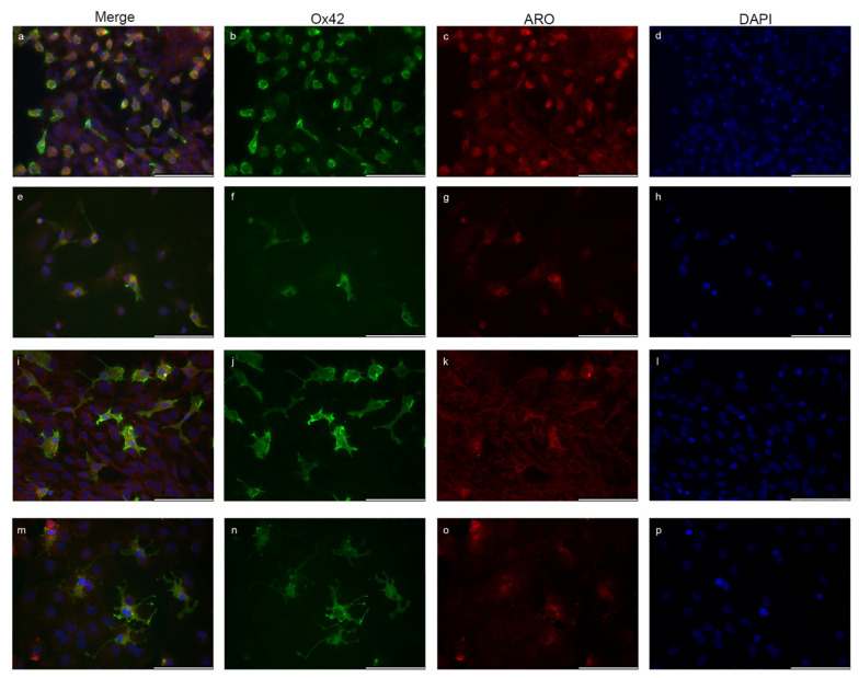
Estrogens regulate a variety of neuroendocrine, reproductive and also non-reproductive brain functions. Estradiol biosynthesis in the central nervous system (CNS) is catalyzed by the enzyme aromatase, which is expressed in several brain regions by neurons, astrocytes and microglia. In this study, we performed a complex fluorescent immunocytochemical analysis which revealed that aromatase is colocalized with the nuclear stain in glial fibrillary acidic protein (GFAP) positive astrocytes in cell cultures. Confocal immunofluorescent Z-stack scanning analysis confirmed the colocalization of aromatase with the nuclear DAPI signal. Nuclear aromatase was also detectable in the S100β positive astrocyte subpopulation. When the nuclear aromatase signal was present, estrogen receptor alpha was also abundant in the nucleus. Immunostaining of frozen brain tissue sections showed that the nuclear colocalization of the enzyme in GFAP-positive astrocytes is also detectable in the adult rat brain. CD11b/c labelled microglial cells express aromatase, but the immunopositive signal was distributed only in the cytoplasm both in the ramified and amoeboid microglial forms. Immunostaining of rat ovarian tissue sections and human granulosa cells revealed that aromatase was present only in the cytoplasm. This novel observation suggests a new unique mechanism in astrocytes that may regulate certain CNS functions via estradiol production.

Keywords: aromatase; astrocyte; central nervous system; estrogen; estrogen receptor alpha; microglia.


Conflict of interest statement

The authors declare no conflict of interest.

Figures

Figure 1
Figure 1
Aromatase (Aro) expression in astrocytes in glia-enriched subcultures. Cells were isolated from P0–P5 newborn rats. Aro (red) is strongly expressed in GFAP-positive astrocytes (green) and colocalizes with the nuclear staining (DAPI, blue). Fibrous astrocytes with small somata and long, thin unbranched processes (ah) show strong nuclear Aro expression (a,c,d, white arrowheads). Cytoplasmic Aro expression is weaker, and the long thin processes do not show Aro immunopositivity. Protoplasmic astrocytes (ip) with bigger somata and short, thick and frequently branched processes also display nuclear Aro expression (i,k,l, white arrowheads). Weak immunopositivity is detectable in the processes. The nuclear Aro signal distributes evenly or in a dotted pattern (c,k, white arrowheads) or strongly concentrates to certain parts of the nucleus (g) in both astrocyte subtypes. (White arrowheads indicate a selected representative cell and its nucleus with the Aro signal). Scale bar: 100 μm.
Figure 2
Figure 2
Confocal immunofluorescent analysis of GFAP positive astrocytes. Z-stack scanning analysis confirms Aro (red) immunopositivity in the nuclear layer. One representative cell with GFAP (green) and nuclear Aro immunopositivity (a) is selected for Z-stack scanning (c). The selected astrocyte shows dotted nuclear Aro positivity (b). Orthogonal scales from Z3 layer prove that the Aro signal originates from the nucleus (b) as the immunopositive dots colocalize with the nuclear signal (blue). Magnification: 60×.
Figure 3
Figure 3
Nuclear colocalization of Aro in S100β positive cells from P0–P5 newborn rats. The three individual sets of pictures (al) show that Aro (red) is also expressed in S100β-positive astrocytes (green) in glial cell culture. Aro immunopositivity is detectable in the close perinuclear cytoplasmic area (c,g,k; yellow arrowheads) and also colocalizes (a,c,e,g,i,k; white arrowheads) with the nuclear signal (blue). Scale bar: 100 μm.
Figure 4
Figure 4
Quantitative analysis of aromatase localization in astrocytes. Analysis of mean fluorescent intensity revealed that Aro intensity is significantly stronger in the nucleus than in the cytoplasm in different astrocyte subtypes. GFAP-positive fibrous astrocytes showed stronger nuclear Aro intensity (36.36 ± 7.7; n = 25) than GFAP-positive protoplasmic astrocytes (32.25 ± 9.18; n = 29). Nuclear Aro intensity in the S100b-positive subtype (21.72 ± 4.98; n = 24) was significantly lower than in the GFAP-positive subtype. Data are presented in mean ± SD. * p < 0.05, *** p < 0.001, ### p < 0.001. Asterisks indicate the significance between the nucleus and cytoplasm within each group. Pound (###) indicates the significantly lower intensity of nuclear Aro between GFAP and S100b positive astrocyte subtypes.
Figure 5
Figure 5
Localization of aromatase in astrocytes in adult rat brain sections. Nuclear (nucleus: blue) appearance of Aro enzyme (red; c,g,k,o; yellow arrowheads) in GFAP-labelled (green) astrocytes (a,b,e,f,i,j,m,n; white arrowheads) is also detectable in frozen cortical tissue sections from male (ah) and female (ip) rats. Interestingly, the Aro immunopositive signal in the adult cortical astrocytes is abundant in the processes (c,g,k,o; green arrowheads), while astrocytes derived from newborn rats do not show an Aro signal in the branches. Quantitative analysis of total GFAP-positive astrocytes and nuclear Aro immunopositive astrocytes (q) in 10 randomly selected microscopic fields of different frozen sections from both sexes showed that the abundance of nuclear Aro immunopositive GFAP-labelled astrocytes is about 20% in both males (19.79%; n = 197) and females (20.13%; n = 154). Scale bar: 100 μm.
Figure 6
Figure 6
Localization of aromatase in microglia cells. CD11b/c (Ox42)-labelled microglia cells (green) isolated from newborn rats show Aro (red) expression, but the signal is detectable exclusively in the cytoplasm. Microglia cells with typical ameboid/activated morphology (ah) show stronger Aro expression compared with the resting/ramified forms (ip), where Aro immunopositivity is much weaker. Aro signal is weak or undetectable in the branches of ramified microglia cells. Quantitative analysis of the fluorescent intensity confirmed that the Aro signal is significantly higher in the cytoplasm of ameboid cells (18.64 ± 2.81; n = 15) than in ramified cells (9.98 ± 1.35; n = 15) (r). Although Aro is not presented in the nucleus of microglia cells, a low level of fluorescent intensity was detectable in the nuclear area due to the overprojection of staining of neighbouring cells from the upper and lower cellular layers and/or the overlapping of cytoplasmic signal, but its level was significantly lower (9.71 ± 2.31; n = 45) than the cytoplasmic Aro intensity (14.67 ± 4.68; n = 45). The analysis of mean fluorescent intensity also revealed that the level of Aro immunopositivity in the cytoplasm is similar in astrocytes (14.20 ± 4.9; n = 54) and microglia cells (q). Data are presented in mean ± SD. ** p < 0.01, *** p < 0.001, ### p < 0.001. Asterisks indicate the significance between microglia cells. Pound (###) indicates the significance between astrocytes and microglia. Scale bar: 100 μm.
Figure 6
Figure 6
Localization of aromatase in microglia cells. CD11b/c (Ox42)-labelled microglia cells (green) isolated from newborn rats show Aro (red) expression, but the signal is detectable exclusively in the cytoplasm. Microglia cells with typical ameboid/activated morphology (ah) show stronger Aro expression compared with the resting/ramified forms (ip), where Aro immunopositivity is much weaker. Aro signal is weak or undetectable in the branches of ramified microglia cells. Quantitative analysis of the fluorescent intensity confirmed that the Aro signal is significantly higher in the cytoplasm of ameboid cells (18.64 ± 2.81; n = 15) than in ramified cells (9.98 ± 1.35; n = 15) (r). Although Aro is not presented in the nucleus of microglia cells, a low level of fluorescent intensity was detectable in the nuclear area due to the overprojection of staining of neighbouring cells from the upper and lower cellular layers and/or the overlapping of cytoplasmic signal, but its level was significantly lower (9.71 ± 2.31; n = 45) than the cytoplasmic Aro intensity (14.67 ± 4.68; n = 45). The analysis of mean fluorescent intensity also revealed that the level of Aro immunopositivity in the cytoplasm is similar in astrocytes (14.20 ± 4.9; n = 54) and microglia cells (q). Data are presented in mean ± SD. ** p < 0.01, *** p < 0.001, ### p < 0.001. Asterisks indicate the significance between microglia cells. Pound (###) indicates the significance between astrocytes and microglia. Scale bar: 100 μm.
Figure 7
Figure 7
ERα is strongly represented in astrocytes. In GFAP (red)-labelled astrocytes (a,b,e,f,i,j; white arrowheads), ERα (green) is heavily expressed (c,g,k). The ERα signal in fibrous astrocytes shows strong and predominantly perinuclear localization in the cytoplasm (c,g,k; blue arrowheads), but immunopositive dots are also detectable in the processes (c; orange arrowheads). Nuclear ERα signals are represented as strong, unevenly distributed immunopositive dots or as a weak but even signal (c,g,k; yellow arrowheads). Nuclei are stained with blue (d,h,l). Scale bar: 100 μm.
Figure 8
Figure 8
Nuclear aromatase expression overlaps with nuclear ERα signal. Double immunostaining with ERα (green) and Aro (red) in glia culture (al) shows that nuclear Aro signals colocalize with nuclear ERα (a,e,i; white arrowheads). Both Aro and ERα are strongly represented in the nucleus (b,c,f,g,j,k; yellow arrowheads) and also in the cytoplasm (b,c,f,g,j,k; blue arrowheads). ERα positive dots are also detectable in the processes (b,f,j; orange arrowheads). Scale bar: 100 μm.
Figure 9
Figure 9
Localization of aromatase in the rat ovary and in human granulosa cells. Immunostaining of rat ovarian tissue sections is presented in images (af). Immunohistochemistry revealed that Aro (red) is strongly expressed in the whole ovarian tissue during the oestrus cycle but does not colocalize with the nuclear signal (blue). Results from Aro-stained human granulosa cells (red) corroborate the immunohistochemical findings as Aro immunopositivity is abundant in granulosa cells but only in the cytoplasm (gl). Scale bar: 100 μm.
Figure 10
Figure 10
The simplified structure of human aromatase (Aro) protein with important conserved regions shown in red (based on Di Nardo et al. [8]). Blue lines indicate which parts of the human Aro protein were used to produce antibodies for Aro detection in CNS studies (Table 1). Green line indicates a mouse Aro peptide to generate an anti-mouse Aro antibody. Co: commercially available antibodies; In-h: antibodies made and validated in-house; * the Novus Aro antibody used in this study.

Similar articles

Cited by

References

    1. Garcia-Ovejero D., Azcoitia I., Doncarlos L.L., Melcangi R.C., Garcia-Segura L.M. Glia-neuron crosstalk in the neuroprotective mechanisms of sex steroid hormones. Brain Res. Rev. 2005;48:273–286. doi: 10.1016/j.brainresrev.2004.12.018. - DOI - PubMed
    1. Azcoitia I., Sierra A., Veiga S., Honda S., Harada N., Garcia-Segura L.M. Brain aromatase is neuroprotective. J. Neurobiol. 2001;47:318–329. doi: 10.1002/neu.1038. - DOI - PubMed
    1. Saldanha C.J., Duncan K.A., Walters B.J. Neuroprotective actions of brain aromatase. Front. Neuroendocrinol. 2009;30:106–118. doi: 10.1016/j.yfrne.2009.04.016. - DOI - PMC - PubMed
    1. Krentzel A.A., Willett J.A., Johnson A.G., Meitzen J. Estrogen receptor alpha, G-protein coupled estrogen receptor 1, and aromatase: Developmental, sex, and region-specific differences across the rat caudate-putamen, nucleus accumbens core and shell. J. Comp. Neurol. 2020;529:786–801. doi: 10.1002/cne.24978. - DOI - PMC - PubMed
    1. Villa A., Vegeto E., Poletti A., Maggi A. Estrogens, Neuroinflammation, and Neurodegeneration. Endocr. Rev. 2016;37:372–402. doi: 10.1210/er.2016-1007. - DOI - PMC - PubMed