. 2022 Aug 11;23(16):8962.
doi: 10.3390/ijms23168962.

(-)-Epicatechin Is a Biased Ligand of Apelin Receptor

Affiliations

(-)-Epicatechin Is a Biased Ligand of Apelin Receptor

Andrés Portilla-Martínez et al. Int J Mol Sci. .

Abstract

(-)-Epicatechin (EC) is part of a large family of biomolecules called flavonoids and is widely distributed in the plant kingdom. Several studies have shown the beneficial effects of EC consumption. Many of these reported effects are exerted by activating the signaling pathways associated with the activation of two specific receptors: the G protein-coupled estrogen receptor (GPER), a transmembrane receptor, and the pregnane X receptor (PXR), which is a nuclear receptor. However, the effects of EC are so diverse that these two receptors cannot describe the complete phenomenon. The apelin receptor or APLNR is classified within the G protein-coupled receptor (GPCR) family, and is capable of activating the G protein canonical pathways and the β-arrestin transducer, which participates in the phenomenon of receptor desensitization and internalization. β-arrestin gained interest in selective pharmacology and mediators of the so-called "biased agonism". With molecular dynamics (MD) and in vitro assays, we demonstrate how EC can recruit the β-arrestin in the active conformation of the APLN receptor acting as a biased agonist.

Keywords: (-)-epicatechin; APLNR; bias ligand; molecular dynamics; β-arrestin.


Conflict of interest statement

F.V. is the founder and stockholder of Epirium Inc., and G.C. is a stockholder of Epirium Inc. The rest of the authors declare no conflict of interest.

Figures

Figure 1
Figure 1
Acute effects (30 min) of EC on upstream Akt activation and the inhibition of these effects with G15 (GPER antagonist), ML221 (APLNR antagonist), and the combination of both. Each Western blot is representative of three independent experiments. Data are expressed as mean ± SD (n = 3). * = p < 0.05, NS (nonsignificant).
Figure 2
Figure 2
(A): Nonlinear regression with the specific binding with hillslope model for apelin-13, (-)-epicatechin (EC), and the mixture with its antagonist ML221. Relative luminescence units (RLU) are plotted on the y-axis, and data are expressed as mean S.E.; (B) dose–response curve of normalized data taking the EC effect as 10%.
Figure 3
Figure 3
(A) (-)-Epicatechin and (B) CMF-019 molecular structures.
Figure 4
Figure 4
In the four images, we present the binding site and the interactions of the molecular docking results. (A) CMF-019 binds to the iAPLNR receptor and shows eight hydrophobic interactions and one polar interaction, of which it is worth highlighting two π-π interactions with the Phe291 residue and the hydrogen bond with the Arg168 residue. (B) The CMF-019 binds to the aAPLNR receptor and establishes eight hydrophobic interactions, two electrostatic interactions, and one ionic, of which we can highlight two π-π with the Phe291 residue; two hydrogen bonds, one with Tyr88 and one with Tyr264; and finally, the formation of a π-cation with residue Arg168. (C) EC in the inactive conformation of the receptor (iAPLNR) establishes few interactions in contrast to the other three assays, counting only two interactions of a hydrophobic nature with residues Pro292 and Tyr299. (D) On the other hand, the EC in the active conformation of the receptor (aAPLNR) establishes four interactions, three hydrophobic and one electrostatic, of which it is worth highlighting the hydrogen bond with Tyr182 and the π-π with Trp85.APJ = APLNR. Images were made with Discovery Studio [39].
Figure 5
Figure 5
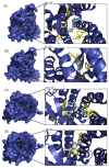
On the left side of each image, the APLN receptor seen from above is shown in a surface representation, and within the cavity (binding site) is the corresponding ligand. A close-up of the binding site is shown to the right of each image, with the amino acid residues in yellow and the ligand in cyan blue. (A) The binding site of the CMF-019 in the inactive APLNR conformation. (B) The binding site of the (-)-epicatechin in the inactive APLN conformation. (C) The binding site of the CMF-019 in the active APLNR conformation. (D) The binding site of the (-)-epicatechin in the active APLNR conformation.
Figure 6
Figure 6
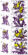
On the left side of each model, the APJ receptor seen from the side is represented in 3D, and on the right, the same receptor, but seen from above. Yellow shows the surface of the residues with which the ligand is in contact, and the arrows indicate the transmembrane domain (TM) to which the residue corresponds. (A) The interaction of CMF-019 with the receptor in its inactive conformation: it is observed how the ligand has contact with TM2, 3, 4, 6, and 7. (B) The interaction of (-)-epicatechin with the receptor in its inactive conformation: it is observed how it only has contact with the TM7. (C) The interaction of CMF-019 with the receptor in its active conformation: it is observed how the ligand has contact with TM2, 3, 4, 6, and 7, and with the extracellular loop (ECL) 2. (D) The interaction of (-)-epicatechin with the receptor in its active conformation: it is observed how it has contact with the TM2, 3 and 7, and with the ECL2.
Figure 7
Figure 7
APLNR in both conformations superimposed. Active on purple mesh and inactive on blue surface.
Figure 8
Figure 8
The RMSD plots were obtained from a 100 ns MD simulation. On the ordinate axis, the variation in distance is shown in nanometers and on the abscissa axis, the trajectory time is shown in nanoseconds. In all quadrants, we can see the movement of the APLN receptor alone (black line), the APLN receptor in complex with a ligand (red line), the CMF ligand only in (A,B) (purple line), and the EC ligand only in (C,D) (green line). iAPLNR = inactive APLNR; aAPLNR = active APLNR; EC = (-)-epicatechin; CMF = CMF-019; APJ = APLNR; black line = receptors MD simulation; red line = receptor in complex with a ligand; green = EC within the receptor; purple line = CMF-019 within the receptor; ns = nanoseconds; nm = nanometers.
Figure 9
Figure 9
The H-bond plot shows the evolution of all hydrogen bonds formed by (-)-epicatechin (EC) and CMF-019 (CMF) in complex with the iAPLNR and aAPLNR, during a 100 ns MD simulation. For both graphs, on the ordinate axis, we see the number of hydrogen bonds, and on the abscissa axis, we have the simulation time expressed in nanoseconds. (A) The CMF can form up to six hydrogen bonds in the active conformation, and five in the inactive one of the APLN receptor during the entire trajectory, with an average of approximately three hydrogen bonds for both conformations. (B) EC manages to form up to five hydrogen bonds in the active conformations and up to six hydrogen bonds in the inactive conformation. For the active conformation of the receptor (aAPLNR), the hydrogen bonds begin to disappear in the last ~30 ns of the simulation, until only one is formed. This is because in the presence of EC, the APLNR modifies its conformation after a period of time—the same phenomenon that we observed with the RMSD of the EC + aAPLNR complex. ns = nanoseconds.
Figure 10
Figure 10
The H-bond plot shows the evolution of the hydrogen bonds formed by the TM5 Y221 residue and the TM7 Y309 residue. (A) When the receptor is alone (aAPLNR—purple lines), we see how these two residues form the hydrogen bond in practically the entire trajectory. However, when CMF-109 (CMF) binds, it induces a conformational change in the receptor that causes a gap between the TM5 and TM7, thus preventing the formation of the hydrogen bond. (B) On the other hand, when (-)-epicatechin (EC) is inside, it allows the receptor to maintain the spatial arrangement so that these two tyrosine residues are close enough to form the hydrogen bond at least until nanosecond ~75, which is when CE begins to induce a conformational change in the receptor, as we had observed in the RMSD and protein–ligand hydrogen bond formation analysis. APJ = APLNR; ns = nanoseconds.
Figure 11
Figure 11
In the images, the aAPLNR and the same receptor residue Arg127 are shown in green. Cyan blue shows the G protein and the same protein residue, Tyr356. (A) It can be seen how, in the simulation, the aAPLNR in complex with CMF-019 is at a sufficient distance to allow the G protein to enter the cavity. (B) On the other hand, the aAPLNR in complex with the EC is in such a conformation that when placing the G protein, the Tyr356 residue collides spatially with the Arg127 residue of the receptor, which would indicate that the G protein cannot be attached in the closed cavity.
Figure 12
Figure 12
Acute effects (30 min) of apelin−13 and (-)-epicatechin (EC) on upstream Akt activation. These effects were blocked by the β-arrestin inhibitor, Barbadin, as shown in the representative blots (each Western blot is representative of three independent experiments). Data are expressed as mean ± SD (n = 3). * = p < 0.05.

Similar articles

Cited by

References

    1. Taub P.R., Ramirez-Sanchez I., Ciaraldi T.P., Gonzalez-Basurto S., Coral-Vazquez R., Perkins G., Hogan M., Maisel A.S., Henry R.R., Ceballos G., et al. Perturbations in skeletal muscle sarcomere structure in patients with heart failure and Type 2 diabetes: Restorative effects of (-)-epicatechinrich cocoa. Clin. Sci. 2013;125:383–389. doi: 10.1042/CS20130023. - DOI - PubMed
    1. Ramirez-Sanchez I., Taub P.R., Ciaraldi T.P., Nogueira L., Coe T., Perkins G., Hogan M., Maisel A.S., Henry R.R., Ceballos G., et al. (-)-Epicatechin rich cocoa mediated modulation of oxidative stress regulators in skeletal muscle of heart failure and type 2 diabetes patients. Int. J. Cardiol. 2013;168:3982–3990. doi: 10.1016/j.ijcard.2013.06.089. - DOI - PMC - PubMed
    1. Nogueira L., Ramirez-Sanchez I., Perkins G.A., Murphy A., Taub P.R., Ceballos G., Villarreal F.J., Hogan M.C., Malek M.H. (-)-Epicatechin enhances fatigue resistance and oxidative capacity in mouse muscle. J. Physiol. 2011;589:4615–4631. doi: 10.1113/jphysiol.2011.209924. - DOI - PMC - PubMed
    1. Gutiérrez-Salmeán G., Ortiz-Vilchis P., Vacaseydel C.M., Garduño-Siciliano L., Chamorro-Cevallos G., Meaney E., Villafaña S., Villarreal F., Ceballos G., Ramírez-Sánchez I. Effects of (-)-epicatechin on a diet-induced rat model of cardiometabolic risk factors. Eur. J. Pharmacol. 2014;728:24–30. doi: 10.1016/j.ejphar.2014.01.053. - DOI - PubMed
    1. Moreno-Ulloa A., Miranda-Cervantes A., Licea-Navarro A., Mansour C., Beltrán-Partida E., Donis-Maturano L., de la Herrán H.C.D., Villarreal F., Álvarez-Delgado C. (-)-Epicatechin Stimulates Mitochondrial Biogenesis and Cell Growth in C2C12 Myotubes via the G-Protein Coupled Estrogen Receptor. Eur. J. Pharmacol. 2018;822:95. doi: 10.1016/j.ejphar.2018.01.014. - DOI - PMC - PubMed

MeSH terms

LinkOut - more resources