Review
. 2022 Aug 12;23(16):8988.
doi: 10.3390/ijms23168988.

Studying Chromatin Epigenetics with Fluorescence Microscopy

Affiliations
Review

Studying Chromatin Epigenetics with Fluorescence Microscopy

Afanasii I Stepanov et al. Int J Mol Sci. .

Abstract

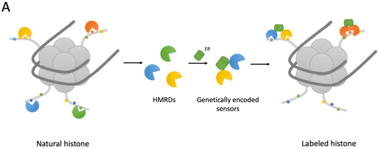
Epigenetic modifications of histones (methylation, acetylation, phosphorylation, etc.) are of great importance in determining the functional state of chromatin. Changes in epigenome underlay all basic biological processes, such as cell division, differentiation, aging, and cancerous transformation. Post-translational histone modifications are mainly studied by immunoprecipitation with high-throughput sequencing (ChIP-Seq). It enables an accurate profiling of target modifications along the genome, but suffers from the high cost of analysis and the inability to work with living cells. Fluorescence microscopy represents an attractive complementary approach to characterize epigenetics. It can be applied to both live and fixed cells, easily compatible with high-throughput screening, and provide access to rich spatial information down to the single cell level. In this review, we discuss various fluorescent probes for histone modification detection. Various types of live-cell imaging epigenetic sensors suitable for conventional as well as super-resolution fluorescence microscopy are described. We also focus on problems and future perspectives in the development of fluorescent probes for epigenetics.

Keywords: epigenetics; fluorescent proteins; genetically encoded probes; histone modification.


Conflict of interest statement

The authors declare no conflict of interest.

Figures

Figure 2
Figure 2
Schematic representation of the antibody-derived probes for histone modification imaging [16,17,18,19,20,21,22,23,24,26].
Figure 5
Figure 5
Reader domain-based sensors for live-cell imaging. (A) General design of reader domain-based sensors. (B) Domain structures of reporters described below [79,80,81].
Figure 5
Figure 5
Reader domain-based sensors for live-cell imaging. (A) General design of reader domain-based sensors. (B) Domain structures of reporters described below [79,80,81].
Figure 1
Figure 1
Schematic outline of MIEL. Cells of interest (e.g., cells at different stages of differentiation and cells after drug treatment) are stained using antibodies against target epigenetic modification(s). The obtained intranuclear patterns (landscapes) of distribution of histone modifications undergo computer analysis (texture features extraction). Cells are compared and classified using the multiparametric Euclidean distances between them.
Figure 3
Figure 3
Genetically encoded FRET sensors for live-cell imaging. (A) General design of FRET sensors. (B,C) Domain structures of FP-based and scFv-based FRET sensors, respectively. Here, and in analogous schemes, below protein parts are not to scale [36,37,38,39,40,41,42,43,44,45].
Figure 4
Figure 4
BiFC sensors for live-cell imaging. (A) General design of BiFC sensors. (B,C) Domain structures of FP-based and scFv-based BiFC sensors, respectively [49,53,54,58,63].
Figure 6
Figure 6
Model to illustrate the spatial organization of the chromatin environment at interphase and mitotic phases. Three distinct structural groups of active histone acetylation, active histone methylation, and repressive histone methylation, as well as their spatial relationship with active transcription machinery are shown. Modified from [91].
Figure 7
Figure 7
Visualizing spatial epigenomics using localization-based super-resolution microscopy. Two-color super-resolution images of chromosomes (green) and histone marks (magenta) via immunostained with anti-SYCP3 (Alexa 555) and anti-histone modifications (Alexa 488). Adapted with permission from [94], 2022, John Wiley and Sons.

References

    1. Buenrostro J.D., Wu B., Litzenburger U.M., Ruff D., Gonzales M.L., Snyder M.P., Chang H.Y., Greenleaf W.J. Single-Cell Chromatin Accessibility Reveals Principles of Regulatory Variation. Nature. 2015;523:486–490. doi: 10.1038/nature14590. - DOI - PMC - PubMed
    1. Barros-Silva D., Marques C.J., Henrique R., Jerónimo C. Profiling DNA Methylation Based on Next-Generation Sequencing Approaches: New Insights and Clinical Applications. Genes. 2018;9:429. doi: 10.3390/genes9090429. - DOI - PMC - PubMed
    1. Karemaker I.D., Vermeulen M. Single-Cell DNA Methylation Profiling: Technologies and Biological Applications. Trends Biotechnol. 2018;36:952–965. doi: 10.1016/j.tibtech.2018.04.002. - DOI - PubMed
    1. Kurdyukov S., Bullock M. DNA Methylation Analysis: Choosing the Right Method. Biology. 2016;5:3. doi: 10.3390/biology5010003. - DOI - PMC - PubMed
    1. Dixon J.R., Selvaraj S., Yue F., Kim A., Li Y., Shen Y., Hu M., Liu J.S., Ren B. Topological Domains in Mammalian Genomes Identified by Analysis of Chromatin Interactions. Nature. 2012;485:376–380. doi: 10.1038/nature11082. - DOI - PMC - PubMed

LinkOut - more resources