Review
. 2022 Aug 12;23(16):9013.
doi: 10.3390/ijms23169013.

The Relevance of Crystal Forms in the Pharmaceutical Field: Sword of Damocles or Innovation Tools?

Affiliations
Review

The Relevance of Crystal Forms in the Pharmaceutical Field: Sword of Damocles or Innovation Tools?

Dario Braga et al. Int J Mol Sci. .

Abstract

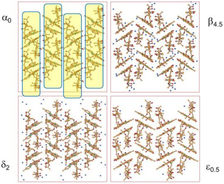
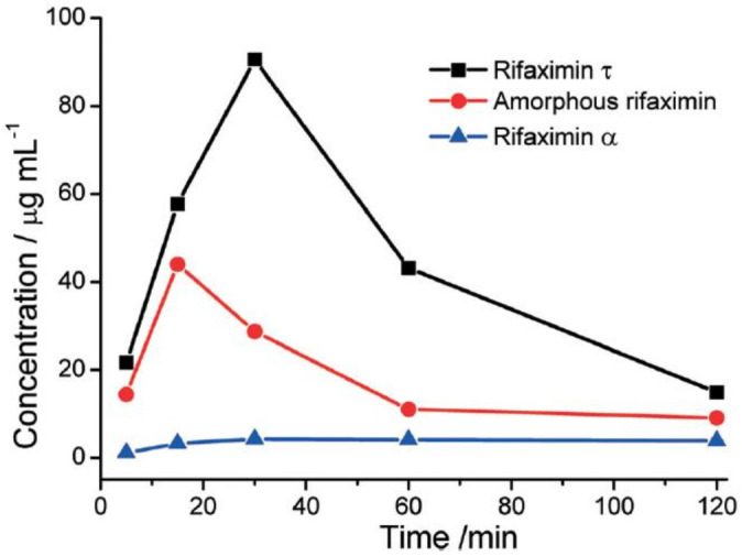
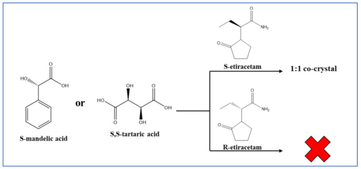
This review is aimed to provide to an "educated but non-expert" readership and an overview of the scientific, commercial, and ethical importance of investigating the crystalline forms (polymorphs, hydrates, and co-crystals) of active pharmaceutical ingredients (API). The existence of multiple crystal forms of an API is relevant not only for the selection of the best solid material to carry through the various stages of drug development, including the choice of dosage and of excipients suitable for drug development and marketing, but also in terms of intellectual property protection and/or extension. This is because the physico-chemical properties, such as solubility, dissolution rate, thermal stability, processability, etc., of the solid API may depend, sometimes dramatically, on the crystal form, with important implications on the drug's ultimate efficacy. This review will recount how the scientific community and the pharmaceutical industry learned from the catastrophic consequences of the appearance of new, more stable, and unsuspected crystal forms. The relevant aspects of hydrates, the most common pharmaceutical solid solvates, and of co-crystals, the association of two or more solid components in the same crystalline materials, will also be discussed. Examples will be provided of how to tackle multiple crystal forms with screening protocols and theoretical approaches, and ultimately how to turn into discovery and innovation the purposed preparation of new crystalline forms of an API.

Keywords: co-crystals of active pharmaceuticals; crystal polymorphism; hydrates.


Conflict of interest statement

The authors declare no conflict of interest.

Figures

Figure 26
Figure 26
(a) The effect of co-crystallization on the dissolution rate for co-crystals of hesperetin with picolinic acid, nicotinamide, and caffeine. (b) Equilibrium solubility (24 h) of hesperetin, HESP-PICO, HESP-NICO, and HESP-CAFF. Reprinted with permission from Ref. [89]. 2017, American Chemical Society.
Figure 1
Figure 1
Differences in shape and color between the first ten (out of 13) discovered polymorphs of ROY (ROY = red, orange, yellow polymorphs of 5-methyl-2-[(2-nitrophenyl) amino]-3-thiophene carbonitrile). Reprinted with permission from Ref. [21]. 2010, American Chemical Society.
Figure 2
Figure 2
E/T diagrams, with G and H vs. temperature in the enantiotropic (left) and monotropic cases (right).
Figure 3
Figure 3
Hydrogen bonded chains in paracetamol Form I (top, refcode HXACAN04) and Form II (bottom, refcode HXACAN21).
Figure 4
Figure 4
Crystal habits and shapes of the two polymorphs of paracetamol. The outcome of the crystallization can be changed by doping the crystallization with metacetamol. Reprinted with permission from Ref. [28]. 2020, American Chemical Society.
Figure 5
Figure 5
Crystal packing of chloramphenicol palmitate Form B (view down the crystallographic c-axis).
Figure 6
Figure 6
E/T (energy vs. temperature) diagram showing the complex phase relationship between the three unsolvated, non-hygroscopic crystalline forms, designated as form A, form B, and form C of Bitopertin. Figure adapted from [32].
Figure 7
Figure 7
Schematic representation of the concepts of “conformation change” and “conformational adjustment” in a PES (potential energy surface) profile. Reprinted with permission from Ref. [33]. 2014, American Chemical Society.
Figure 8
Figure 8
(Top) The α and β forms of L-glutamic acid and a comparison of the hydrogen bonded chains in the α (middle) and β (bottom) polymorphs.
Figure 9
Figure 9
(Top): keto/enol tautomerism of barbituric acid; (bottom): crystal structure at room temperature of the enol form.
Figure 10
Figure 10
(a) Keto/enol tautomerism of thiobarbituric acid and (b) the crystal structure of the most stable form at room temperature, showing the presence of both the keto and enol forms [37].
Figure 11
Figure 11
The hydrogen bonding networks in crystals of Ritonavir form I (left) and form II (right) [38].
Figure 12
Figure 12
The appearance of the stable crystalline form of Rotigotine on the patches used for administering the drug. Reprinted with permission from Ref. [43]. 2015, Elsevier.
Figure 13
Figure 13
A flow-chart showing how polymorph screening ought to be associated with the API selection, before entering trial stage, and the quality control process, once the API is on the market.
Figure 14
Figure 14
A representation of the types of association of a solvent with a crystalline solid.
Figure 15
Figure 15
(Top): packing comparison and interconversion process between anhydrous and trihydrate ampicillin (refcodes AMCILL and AMPCIH01, respectively). (Bottom): comparison between the powder diffractograms calculated on the basis of the single-crystal data, showing the difference between the two patterns.
Figure 16
Figure 16
(Top): The structures of the anhydrate ((a), refcode BAPLOT01) and of the monohydrate ((b), refcode THEOPH01) crystals of theophylline. The anhydrate is obtained from a mixture of an organic solvent and water at low water activity. (Bottom): comparison of the the powder diffractograms calculated on the basis of the single-crystal data, showing the difference between the two patterns.
Figure 16
Figure 16
(Top): The structures of the anhydrate ((a), refcode BAPLOT01) and of the monohydrate ((b), refcode THEOPH01) crystals of theophylline. The anhydrate is obtained from a mixture of an organic solvent and water at low water activity. (Bottom): comparison of the the powder diffractograms calculated on the basis of the single-crystal data, showing the difference between the two patterns.
Figure 17
Figure 17
(a) The intricate hydration/dehydration phase relationship between the various forms of Rifaximin, (b) the powder X-ray diffraction patterns five forms of Rifaximin, (c) dissolution rates of Rifaximin hydrates at 250 (left) and 100 (right) rpm of mixing rate. Time vs. absorbance at 293 nm. Reproduced with permission from [63,64].
Figure 18
Figure 18
The single crystal to single crystal form β → form α transformation. The red and green lines evidence the change in the monoclinic β-angle from 91° in Rifaximin form α to 110° in Rifaximin form β. Reprinted with permission from Ref. [63]. 2019, Royal Society of Chemistry.
Figure 19
Figure 19
A comparison of molecular packings in four hydrate forms of the antibiotic Rifaximin. Reproduced with permission from [63].
Figure 20
Figure 20
The solid-state structure of Rifaximin τ, showing the interaction of Rifaximin with transcutol® [67,68]. Reprinted with permission from Ref. [67]. 2019, Royal Society of Chemistry.
Figure 21
Figure 21
Comparison of dissolution rates for Rifaximin τ, amorphous Rifaximin, and Rifaximin α at neutral pH. Reprinted with permission from Ref. [67]. 2019, Royal Society of Chemistry.
Figure 22
Figure 22
Hydrogen bonding pattern in the 1:1 co-crystal of 1-methyl adenine and 1-methyl thymine, first reported in 1963 by Hoogsteen (CSD refcode MTHMAD).
Figure 23
Figure 23
A schematic representation of two polymorphs of a co-crystal of the same API and a conformer.
Figure 24
Figure 24
The acid–amide synthon frequently occurring in the co-crystals of carbamazepine with carboxylic acids.
Figure 25
Figure 25
Packing diagram of the melatonine/pimelic acid co-crystal showing the hydrogen bonding (A). Powder dissolution profiles of melatonine, melatonine-pimelic acid co-crystal, and of a 1:1 physical mixture in PBS (pH 6.8) at 37 °C (B). Adapted with permission from Ref. [87]. 2015, Royal Society of Chemistry.
Figure 27
Figure 27
If RS-resorcylmethylcarbinol (left) and brucine (right) are co-crystallized from methanol, only dextro-resorcylmethyl carbinol forms a co-crystal with brucine [90].
Figure 28
Figure 28
Resolution of optical isomers of 4-amino-p-chlorobutyric acid lactam (Baclofen®) by co-crystallization 4-amino-p-chlorobutyric acid lactam lactam [91].
Figure 29
Figure 29
The 2:1 co-crystal of (R-)-4-amino-p-chlorobutyric acid lactam with (2R,3R)-tartaric acid [91].
Figure 30
Figure 30
The enantioselective co-crystal formation in the case of S-enantiomer of 2-(2-oxopyrrodin-1-yl)butanamide (S-etiracetam) with S-mandelic and S-tartaric acid. The same reaction with R-etiracetam does not lead to co-crystal formation [92].
Figure 31
Figure 31
KUCJAT. The carbons of 3-methylcyclohexane are marked in orange and HCH have been removed for the sake of clarity [95].

Similar articles

Cited by

References

    1. Mitscherlich E. Sur la relation qui existe entre la forme cristalline e le proportions chimiques, I. Memoires sur les arseniates et les phosphates. Ann. Chim. Phys. 1822;19:350–419.
    1. McCrone W.C. Physics and Chemistry of the Organic Solid State. In: Fox D., Labes M.M., Weissenberg A., editors. Physics Today. 2nd ed. Volume 2. Interscience; New York, NY, USA: 1965. 725p
    1. Bernstein J. Polymorphism in Molecular Crystals. 2nd ed. Oxford University Press; Oxford, UK: 2020.
    1. Brittain H.G., editor. Polymorphism in Pharmaceutical Solids. 2nd ed. Informa Healthcare; London, UK, USA: 1999.
    1. Hilfiker R., Von Raumer M., editors; Hilfiker R., Von Raumer M., editors. Polymorphism in the Pharmaceutical Industry: Solid Form and Drug Development. 2nd ed. Wiley-VCH; Weinheim, Germany: 2019.

LinkOut - more resources