Comparative Study
. 2022 Aug 13;23(16):9058.
doi: 10.3390/ijms23169058.

A Comparative Study of the Plasma Chemokine Profile in COVID-19 Patients Infected with Different SARS-CoV-2 Variants

Affiliations
Comparative Study

A Comparative Study of the Plasma Chemokine Profile in COVID-19 Patients Infected with Different SARS-CoV-2 Variants

Zoia R Korobova et al. Int J Mol Sci. .

Abstract

Background: Infection caused by SARS-CoV-2 mostly affects the upper and lower respiratory tracts and causes symptoms ranging from the common cold to pneumonia with acute respiratory distress syndrome. Chemokines are deeply involved in the chemoattraction, proliferation, and activation of immune cells within inflammation. It is crucial to consider that mutations within the virion can potentially affect the clinical course of SARS-CoV-2 infection because disease severity and manifestation vary depending on the genetic variant. Our objective was to measure and assess the different concentrations of chemokines involved in COVID-19 caused by different variants of the virus.

Methods: We used the blood plasma of patients infected with different variants of SARS-CoV-2, i.e., the ancestral Wuhan strain and the Alpha, Delta, and Omicron variants. We measured the concentrations of 11 chemokines in the samples: CCL2/MCP-1, CCL3/MIP-1α, CCL4/MIP-1β, CCL7/MCP-3, CCL11/Eotaxin, CCL22/MDC, CXCL1/GROα, CXCL8/IL-8, CXCL9/MIG, CXCL10/IP-10, and CX3CL1/Fractalkine.

Results: We noted a statistically significant elevation in the concentrations of CCL2/MCP-1, CXCL8/IL-8, and CXCL1/IP-10 independently of the variant, and a drop in the CCL22/MDC concentrations.

Conclusions: The chemokine concentrations varied significantly depending on the viral variant, leading us to infer that mutations in viral proteins play a role in the cellular and molecular mechanisms of immune responses.

Keywords: COVID-19; Omicron; SARS-CoV-2 variants; chemokines; macrophage-derived chemokine.


Conflict of interest statement

The authors declare no conflict of interest.

Figures

Figure 1
Figure 1
A comparison of the concentrations of the chemokines in the plasma of individuals infected with the original strain of SARS-CoV-2 in the acute stage (COVID-19), n = 56, and in healthy donors (HD), n = 51. The concentrations are presented in pg/mL plots representing medians with interquartile ranges. For CXCL10/IP-10, the Y-axis is presented as a logarithmic scale. Only chemokines with a statistically significant difference are presented.
Figure 2
Figure 2
A comparison of the concentrations of the chemokines in the plasma of individuals infected with the Alpha variant of SARS-CoV-2 in the acute stage (COVID-19), n = 95, and in healthy donors (HD), n = 51. The concentrations are presented in pg/mL plots representing medians with interquartile ranges. Only chemokines with a statistically significant difference are presented.
Figure 3
Figure 3
A comparison of the concentrations of the chemokines in the plasma of individuals infected with the Delta variant of SARS-CoV-2 in the acute stage (COVID-19), n = 78 and in healthy donors (HD), n = 51. The concentrations are presented in pg/mL plots representing medians with interquartile ranges. Only chemokines with a statistically significant difference are presented.
Figure 4
Figure 4
A comparison of the concentrations of the chemokines in the plasma of individuals infected with the Omicron variant of SARS-CoV-2 in the acute stage (COVID-19), n = 57, and in the healthy donors (HD), n = 51. The concentrations are presented in pg/mL plots representing medians with interquartile ranges. Only chemokines with a statistically significant difference are presented. Additional data on the differences in the chemokine concentrations for each variant can be found in Supplemental Tables S1–S4.
Figure 5
Figure 5
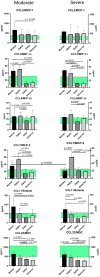
A comparison of the concentrations of C-C chemokines in patients with Wuhan, Alpha, Delta, and Omicron strains, depending on severity. The concentrations are presented in pg/mL (median and interquartile range). The green stripe represents Q25–Q75 of the chemokine concentrations in the samples from healthy donors.
Figure 6
Figure 6
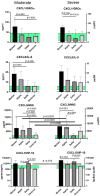
A comparison of the concentrations of C-X-C chemokines in patients with Wuhan, Alpha, Delta, and Omicron strains, depending on severity. The concentrations are presented in pg/mL (median and interquartile range). For CXCL10/IP-10, the concentration axis (Y) is presented as a logarithmic (log10). The green stripe represents Q25–Q75 of the chemokine concentrations in the samples from healthy donors.
Figure 7
Figure 7
A comparison of the concentrations of C-X-3 chemokine Fractalkine in patients with Wuhan, Alpha, Delta, and Omicron strains, depending on severity. The concentrations are presented in pg/mL (median and interquartile range). The green stripe represents Q25–Q75 of the chemokine concentrations in the samples from healthy donors.

References

    1. Wang X.-M., Mannan R., Xiao L., Abdulfatah E., Qiao Y., Farver C., Myers J.L., Zelenka-Wang S., McMurry L., Su F., et al. Characterization of SARS-CoV-2 and host entry factors distribution in a COVID-19 autopsy series. Commun. Med. 2021;24:1. doi: 10.1038/s43856-021-00025-z. - DOI - PMC - PubMed
    1. Ozono S., Zhang Y., Ode H., Sano K., Tan T.S., Imai K., Miyoshi K., Kishigami S., Ueno T., Iwatani Y., et al. SARS-CoV-2 D614G spike mutation increases entry efficiency with enhanced ACE2-binding affinity. Nat. Commun. 2021;12:848. doi: 10.1038/s41467-021-21118-2. - DOI - PMC - PubMed
    1. Samavati L., Uhal B.D. ACE2, Much More Than Just a Receptor for SARS-COV-2. Front. Cell. Infect. Microbiol. 2020;10:317. doi: 10.3389/fcimb.2020.00317. - DOI - PMC - PubMed
    1. Attaway A.H., Scheraga R.G., Bhimraj A., Biehl M., Hatipoğlu U. Severe COVID-19 pneumonia: Pathogenesis and clinical management. BMJ. 2021;372:n436. doi: 10.1136/bmj.n436. - DOI - PubMed
    1. D’Agnillo F., Walters K.-A., Xiao Y., Sheng Z.-M., Scherler K., Park J., Gygli S., Rosas L.A., Sadtler K., Kalish H., et al. Lung epithelial and endothelial damage, loss of tissue repair, inhibition of fibrinolysis, and cellular senescence in fatal COVID-19. Sci. Transl. Med. 2021;13:eabj7790. doi: 10.1126/scitranslmed.abj7790. - DOI - PMC - PubMed

Publication types

Supplementary concepts