. 2022 Aug 15;23(16):9137.
doi: 10.3390/ijms23169137.

Stabilization of the c-Myc Protein via the Modulation of Threonine 58 and Serine 62 Phosphorylation by the Disulfiram/Copper Complex in Oral Cancer Cells

Affiliations

Stabilization of the c-Myc Protein via the Modulation of Threonine 58 and Serine 62 Phosphorylation by the Disulfiram/Copper Complex in Oral Cancer Cells

Gunng-Shinng Chen et al. Int J Mol Sci. .

Abstract

MYC has a short half-life that is tightly regulated through phosphorylation and proteasomal degradation. Many studies have claimed that treatment with disulfiram (DSF) with or without copper ions can cause cancer cell death in a reactive oxygen species (ROS)-dependent manner in cancer cells. Our previous study showed that the levels of c-Myc protein and the phosphorylation of threonine 58 (T58) and serine 62 (S62) increased in DSF-Cu-complex-treated oral epidermoid carcinoma Meng-1 (OECM-1) cells. These abovementioned patterns were suppressed by pretreatment with an ROS scavenger, N-acetyl cysteine. The overexpression of c-Myc failed to induce hypoxia-inducible factor 1α protein expression, which was stabilized by the DSF-Cu complex. In this study, we further examined the regulatory mechanism behind the induction of the c-Myc of the DSF-Cu complex in an OECM-1 cell compared with a Smulow-Glickman (SG) human normal gingival epithelial cell. Our data showed that the downregulation of c-Myc truncated nick and p62 and the induction of the ratio of H3P/H3 and p-ERK/ERK might not be involved in the increase in the amount of c-Myc via the DSF/copper complexes in OECM-1 cells. Combined with the inhibitors for various signaling pathways and cycloheximde treatment, the increase in the amount of c-Myc with the DSF/copper complexes might be mediated through the increase in the stabilities of c-Myc (T58) and c-Myc (S62) proteins in OECM-1 cells. In SG cells, only the c-Myc (T58) protein was stabilized by the DSF-Cu (I and II) complexes. Hence, our findings could provide novel regulatory insights into the phosphorylation-dependent stability of c-Myc in DSF/copper-complex-treated oral squamous cell carcinoma.

Keywords: c-Myc; disulfiram; hypoxia inducible factor 1α; reactive oxygen species.


Conflict of interest statement

The authors declare no conflict of interest.

Figures
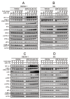
Figure 1
Figure 1
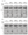
The effects of DSF, copper ions, and DSF/copper complexes on c-Myc and HIF-1α proteins in OECM-1 and SG cells. OECM-1 (A,C) and SG (B,D) cells were treated with the indicated concentrations of copper ions in the absence or presence of DSF for 2 h (A,B) or 4 h (C,D). The cell lysates were subjected to Western blot analysis using antibodies against the indicated proteins. ACTN was a loading protein control. The results are representative of three independent experiments.
Figure 2
Figure 2
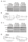
The effects of DSF, copper ions, and DSF/copper complexes on the signaling pathways involved in the c-Myc and HIF-1α proteins in OECM-1 and SG cells. (A) OECM-1 and (B) SG cells were pretreated with the indicated 10 μM of signaling inhibitors for 1 h and then treated with 2 μM of CuCl or 50 μM of CuCl2 plus 0.5 μM of DSF for 2 h (added indicated drugs labeled with +). The cell lysates were subjected to Western blot analysis using antibodies against the indicated proteins. ACTN was a loading protein control. The results are representative of three independent experiments.
Figure 3
Figure 3
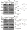
The effects of DSF, copper ions, and DSF/copper complexes on the stability of the c-Myc and p53 proteins in OECM-1 and SG cells. OECM-1 (A) and SG (C) cells were treated with vehicle, 50 μM of CuCl2 plus 0.5 μM of DSF, or 2 μM of CuCl plus 0.5 μM of DSF for 2 h, accompanied by 50 mg/mL CHX for 0, 5, 10, 15, 30, and 60 min. The cell lysates were subjected to Western blot analysis using antibodies against c-Myc and p53 proteins. ACTN was a loading protein control. The protein bands were quantified through pixel density scanning and evaluated using ImageJ, version 1.44a (http://imagej.nih.gov/ij/) (accessed on 1 May 2022). The ratios of c-Myc/ACTN and p53/ACTN were plotted in OECM-1 (B) and SG (D) cells. The results are representative of three independent experiments. The quantified protein bands were compared in cells treated with indicated time of CHX to the 0 h. # p > 0.05, * p < 0.05, ** p < 0.01, and *** p < 0.001.
Figure 4
Figure 4
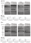
The effects of DSF, copper ions, and DSF/copper complexes on the c-Myc and HIF-1α proteins by NAC and mitoTEMPO in OECM-1 and SG cells. OECM-1 and SG cells were treated with (A) 2 μM of CuCl plus 0.5 μM of DSF and (B) 50 μM of CuCl2 plus 0.5 μM of DSF for 2 h (added indicated drugs labeled with +) in the presence of pretreated NAC, mitoTEMPO, or co-treated NAC with mitoTEMPO for 1 h. The cell lysates were subjected to Western blot analysis using antibodies against the indicated proteins. ACTN was a loading protein control. The results are representative of three independent experiments.
Figure 5
Figure 5
The effects of LiCl on the c-Myc proteins in DSF/copper-complex-treated OECM-1 and SG cells. (A) OECM-1 and (B) SG cells were treated with 0, 5, 10, 20, 40, and 50 mM LiCl for 24 h. The metabolic activity was measured using MTT assays. * p < 0.05, ** p < 0.01, and *** p < 0.001. (C) OECM-1 and (D) SG cells were pretreated with 10 mM LiCl for 1 h and then treated with 50 μM of CuCl2 plus 0.5 μM of DSF or 2 μM of CuCl plus 0.5 μM of DSF for 2 h (added indicated drugs labeled with +). The cell lysates were subjected to Western blot analysis using antibodies against the indicated proteins. ACTN was a loading protein control.
Figure 6
Figure 6
The effects of DSF, copper ions, and DSF/copper complexes on the c-Myc and HIF-1α proteins in LiCl-treated OECM-1 and SG cells. (A) OECM-1 and (C) SG cells were treated with 50 μM of CuCl2 plus 0.5 μM of DSF or 2 μM of CuCl plus 0.5 μM of DSF for 2 h, accompanied by 0, 5, 10, 20, 40, and 50 mM LiCl for 24 h. The cell lysates were subjected to Western blot analysis using antibodies against the indicated proteins. ACTN was a loading protein control. The protein bands (A,C) were quantified through pixel density scanning and evaluated using ImageJ, version 1.44a (http://imagej.nih.gov/ij/) (accessed on 1 May 2022). The ratios of protein/ACTN and pGSK3β/GSK3β were plotted, and red lines are for the DSF/CuCl complexes and black lines for the DSF/CuCl2 complexes in (B) OECM-1 and (D) SG cells.
Figure 7
Figure 7
The effects of DSF, copper ions, and DSF/copper complexes on GSK3β activity for the c-Myc and HIF-1α proteins in OECM-1 and SG cells. (A) OECM-1 and (C) SG cells were treated with 50 μM of CuCl2 plus 0.5 μM of DS or 2 μM of CuCl plus 0.5 μM of DSF for 3 h, accompanied by 50 mg/mL CHX for 0, 5, 10, 15, 30, and 60 min. The cell lysates were subjected to Western blot analysis using antibodies against the indicated proteins. ACTN was a loading protein control. The protein bands (A,C) were quantified through pixel density scanning and evaluated using ImageJ, version 1.44a (http://imagej.nih.gov/ij/) (accessed on 1 May 2022). The ratios of protein/ACTN and pGSK3β/GSK3β are plotted with red lines (vehicle), mint green lines (DSF/CuCl), and black lines (DSF/CuCl2) in (B) OECM-1 and (D) SG cells.

References

    1. Dang C.V., O’Donnell K.A., Zeller K.I., Nguyen T., Osthus R.C., Li F. The c-Myc target gene network. Semin. Cancer Biol. 2006;16:253–264. doi: 10.1016/j.semcancer.2006.07.014. - DOI - PubMed
    1. Meyer N., Penn L.Z. Reflecting on 25 years with MYC. Nat. Rev. Cancer. 2008;8:976–990. doi: 10.1038/nrc2231. - DOI - PubMed
    1. Dang C.V., Resar L.M., Emison E., Kim S., Li Q., Prescott J.E., Wonsey D., Zeller K. Function of the c-Myc oncogenic transcription factor. Exp. Cell Res. 1999;253:63–77. doi: 10.1006/excr.1999.4686. - DOI - PubMed
    1. Adhikary S., Eilers M. Transcriptional regulation and transformation by Myc proteins. Nat. Rev. Mol. Cell Biol. 2005;6:635–645. doi: 10.1038/nrm1703. - DOI - PubMed
    1. Farrell A.S., Sears R.C. MYC degradation. Cold Spring Harb. Perspect. Med. 2014;4:a014365. doi: 10.1101/cshperspect.a014365. - DOI - PMC - PubMed