. 2022 Aug 16;23(16):9214.
doi: 10.3390/ijms23169214.

Structure-Dependent Toxicokinetics of Selected Pyrrolizidine Alkaloids In Vitro

Affiliations

Structure-Dependent Toxicokinetics of Selected Pyrrolizidine Alkaloids In Vitro

Julia Buchmueller et al. Int J Mol Sci. .

Abstract

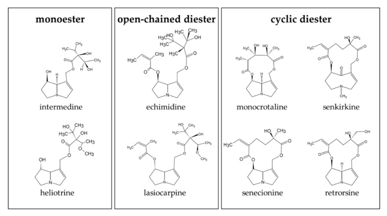
Phytochemicals like pyrrolizidine alkaloids (PAs) can affect the health of humans and animals. PAs can occur for example in tea, honey or herbs. Some PAs are known to be cytotoxic, genotoxic, and carcinogenic. Upon intake of high amounts, hepatotoxic and pneumotoxic effects were observed in humans. This study aims to elucidate different toxicokinetic parameters like the uptake of PAs and their metabolism with in vitro models. We examined the transport rates of differently structured PAs (monoester, open-chained diester, cyclic diester) over a model of the intestinal barrier. After passing the intestinal barrier, PAs reach the liver, where they are metabolized into partially instable electrophilic metabolites interacting with nucleophilic centers. We investigated this process by the usage of human liver, intestinal, and lung microsomal preparations for incubation with different PAs. These results are completed with the detection of apoptosis as indicator for bioactivation of the PAs. Our results show a structure-dependent passage of PAs over the intestinal barrier. PAs are structure-dependently metabolized by liver microsomes and, to a smaller extent, by lung microsomes. The detection of apoptosis of A549 cells treated with lasiocarpine and monocrotaline following bioactivation by human liver or lung microsomes underlines this result. Conclusively, our results help to shape the picture of PA toxicokinetics which could further improve the knowledge of molecular processes leading to observed effects of PAs in vivo.

Keywords: metabolism; pyrrolizidine alkaloids; structure-dependency; uptake.


Conflict of interest statement

The authors declare no conflict of interest.

Figures

Figure 1
Figure 1
Structures of the PAs used in this study. The PAs were chosen to represent the different structures: monoester (intermedine and heliotrine), open-chained diester (echimidine and lasiocarpine) and cyclic diester (monocrotaline, senkirkine, senecionine, and retrorsine).
Figure 2
Figure 2
Transfer rates of structurally different PAs over the differentiated Caco-2 cell monolayer. Caco-2 cells were seeded and differentiated in Transwell inserts and incubated with 0.25 µM of the specific PAs from the apical compartment of the Transwell chamber. At the indicated time points, the basolateral PA amounts were determined with LC-MS/MS. Mean values ± SD of the PA concentrations in the basolateral compartment in % in comparison to the applied PA concentration are shown from three individual experiments.
Figure 3
Figure 3
Reduction of the concentration of PAs after incubation with human intestinal microsomes. 10 µM PAs were incubated with microsomes at 37 °C for up to 4.5 h. At the indicated time points, the remaining PA concentration in comparison to t = 0 h was determined. For each incubation three individual experiments were performed. Mean values ± SD are shown. Statistical differences to t = 0 h were determined with One-Way ANOVA followed by Dunnett’s post hoc test.
Figure 4
Figure 4
Reduction of PA concentrations after incubation with human liver microsomes. 10 µM PAs were incubated with liver microsomes at 37 °C for up to 4.5 h. At the indicated time points, the remaining PA concentration in comparison to t = 0 h was determined. For each incubation three independent experiments were performed. Mean values ± SD are shown. Statistical significance was determined with One-Way ANOVA followed by Dunnett’s post hoc test and is indicated as * p < 0.05, ** p < 0.01.
Figure 5
Figure 5
Reduction of concentration of PAs after incubation with human lung microsomes. 10 µM PAs were incubated with human lung microsomes at 37 °C for up to 4.5 h. At the indicated time points, the remaining PA concentration compared to t = 0 h was determined. For each incubation three independent experiments were performed. Mean values ± SD are shown. Statistical significance was determined with One-Way ANOVA followed by Dunnett’s post hoc test and is indicated as * p < 0.05, ** p < 0.01.
Figure 6
Figure 6
Induction of apoptosis in A549 cells upon incubation with bioactivated PAs: (A) Induction of apoptosis in A549 cells after incubation with 150 µM or 250 µM monocrotaline or lasiocarpine for 4 h + 20 h. As external metabolism system human liver (LiM) or lung (LuM) microsomes were used. Cells were incubated with microsomes (diluted to a final concentration of 2 mg/mL) and PAs for 4 h before washing and incubating for additional 20 h for a total time of 24 h. Induction of apoptosis was measured by flow cytometry by detection of the fluorescence intensities of Annexin-V-FITC and 7-aminoactinomycin (7-AAD). The cells were incubated with medium as negative control and staurosporine as positive control inducing apoptosis or tBOOH as another positive control inducing necrosis. (B) Alteration of Caspase 3 activity in A549 cells upon incubation with 250 µM lasiocarpine or monocrotaline activated priorly with human liver or lung microsomes for 24 h. Cells were incubated with PAs and microsomes for 4 h before washing and further incubation for 20 h. Caspase 3 activity was measured by adding the substrate Ac-DEVD-AFC after lysis of the cells and photometric detection of emission at different time points. For each incubation, three independent experiments were performed. Mean values ± SD are shown. Statistical significance in comparison to the medium control with the respective microsomes but without PAs was determined with One-Way ANOVA followed by Dunnett´s post hoc test and is indicated as * p < 0.05, ** p < 0.01, *** p < 0.001.
Figure 7
Figure 7
Reduction of concentration of lasiocarpine (A) and monocrotaline (B) upon incubation with human supersomes. 10 µM of respective PA were incubated at 37 °C with the CYP supersomes for up to 5 h. At the indicated time points, samples were taken, and the enzymatic reaction was stopped with methanol. The remaining PA concentration was then determined with LC-MS/MS. Three independent experiments were performed for each incubation. Mean values ± SD are shown. Statistical significance was determined with One-Way ANOVA followed by Dunnett´s post hoc test and is indicated as *** p < 0.001.

References

    1. Stegelmeier B., Edgar J., Colegate S., Gardner D., Schoch T., Coulombe R., Molyneux R. Pyrrolizidine alkaloid plants, metabolism and toxicity. J. Nat. Toxins. 1999;8:95–116. doi: 10.2527/jas1988.6692343x. - DOI - PubMed
    1. Wiedenfeld H., Röder E., Bourauel T., Edgar J. Pyrrolizidine Alkaloids: Structure and Toxicity. V&R unipress GmbH; Goettingen, Germany: 2008.
    1. BfR (German Federal Institute for Risk Assessment) Aktualisierte Risikobewertung zu Gehalten an 1,2-ungesättigten Pyrrolizidinalkaloiden (PA) in Lebensmitteln. Stellungnahme 026/2020. BfR; Berlin, Germany: 2020. - DOI
    1. Fu P.P., Xia Q., Lin G., Chou M.W. Pyrrolizidine alkaloids—Genotoxicity, metabolism enzymes, metabolic activation, and mechanisms. Drug Metab. Rev. 2004;36:1–55. doi: 10.1081/DMR-120028426. - DOI - PubMed
    1. Mattocks A., White I. Pyrrolic metabolites from non-toxic pyrrolizidine alkaloids. Nat. New Biol. 1971;231:114–115. doi: 10.1038/newbio231114a0. - DOI - PubMed

LinkOut - more resources