. 2022 Aug 20;23(16):9426.
doi: 10.3390/ijms23169426.

Selective Expansion of NKG2C+ Adaptive NK Cells Using K562 Cells Expressing HLA-E

Affiliations

Selective Expansion of NKG2C+ Adaptive NK Cells Using K562 Cells Expressing HLA-E

Minh-Trang Thi Phan et al. Int J Mol Sci. .

Abstract

Adaptive natural killer (NK) cells expressing self-specific inhibitory killer-cell immunoglobulin-like receptors (KIRs) can be expanded in vivo in response to human cytomegalovirus (HCMV) infection. Developing a method to preferentially expand this subset is essential for effective targeting of allogeneic cancer cells. A previous study developed an in vitro method to generate single KIR+ NK cells for enhanced targeting of the primary acute lymphoblastic leukemia cells; however, the expansion rate was quite low. Here, we present an effective expansion method using genetically modified K562-HLA-E feeder cells for long-term proliferation of adaptive NK cells displaying highly differentiated phenotype and comparable cytotoxicity, CD107a, and interferon-γ (IFN-γ) production. More importantly, our expansion method achieved more than a 10,000-fold expansion of adaptive NK cells after 6 weeks of culture, providing a high yield of alloreactive NK cells for cell therapy against cancer.

Keywords: K562-HLA-E feeder cells; adaptive NK cells; effective expansion; long-term persistence; single KIR NKG2C.


Conflict of interest statement

The authors declare that the research was conducted in the absence of any commercial or financial relationships that could be construed as potential conflicts of interest.

Figures

Figure 1
Figure 1
HLA-E expression of genetically engineered K562 cells. (A) mRNA expression by K562 and K562-HLA-E cells were determined using real-time quantitative polymerase chain reaction (Rq-PCR) with glyceraldehyde-3-phosphate dehydrogenase (GAPDH) as control. (B) HLA-E surface expression was analyzed using fluorescence-activated cell sorting (FACS) with anti-human HLA-E (clone 3D12) in K562 (red) and K562-HLA-E (green) cells.
Figure 2
Figure 2
Purity and fold expansion of expanded natural killer (NK) cells co-cultured with K562 and K562-HLA-E. feeder cells. Freshly isolated peripheral blood mononuclear cells (PBMCs) were co-cultured with γ-irradiated K562 (red) and K562-HLA-E (green) feeder cells in the presence of IL-2 (10 U/mL) at day 0 and IL-2 (100 U/mL) and IL-15 (5 ng/mL) after 1 week. The medium was replaced every 2–3 days along with fresh cytokines. (A) Purity of expanded NK cells was determined by flow cytometry using fluorescein isothio-cyanate (FITC)-conjugated anti-human CD3 and phycoerythrin (PE)-Cy5-conjugated anti-human CD56. (B) Fold expansion of NK cells in the K562 vs. K562-HLA-E added groups was significantly different from day 21 of culture. All data are shown as the mean ± SEM (n = 9 independent experiments; * p < 0.05; ** p < 0.01).
Figure 3
Figure 3
Preferred expansion high frequencies of NKG2C+ natural killer (NK) cells expressing single KIR2DL2/3 by K562-HLA-E in C1C1 donors. (A) Representative fluorescence-activated cell sorting (FACS) plot showing the frequency of NKG2C, NKG2A, KIR2DL1, and KIR2DL2/3 at days 0, 14, and 28 of culture. (B) Percentage expression of NKG2C, KIR2DL2/3, KIR2DL1, and KIR3DL1 on adaptive NK cells (K562-HLA-E) and conventional NK cells (K562) during culture. All data are shown as mean ± SEM (n = 9 independent experiments; * p < 0.05; ** p < 0.01; *** p < 0.001).
Figure 4
Figure 4
Comparison of receptor expression levels of expanded natural killer (NK) cells using K562 and K562-HLA-E cells. (A) Expression of the indicated surface receptors (CD16, NKG2A, CD57, NKp30, NKp46, and NKG2D) on expanded NK cells. Representative histograms are shown in the NK cell subset from the same donor on conventional (K562, red) and adaptive (K562-HLA-E, green) NK cells at day 14 of culture. (B) Differences in the expression levels (percentage of positive) of CD16, NKG2A, CD57, NKp30, NKp46, and NKG2D of conventional (K562) and adaptive (K562-HLA-E) NK cells on day 14 and day 49 of culture (n = 7–9 donors). (C) Differences in the expression levels (mean fluorescence intensity MFI)) of CD16, NKG2A, CD57, NKp30, NKp46, and NKG2D of conventional (K562) and adaptive (K562-HLA-E) NK cells on day 14 and day 49 of culture. All data are shown as mean ± SEM (n = 7–9 independent experiments; * p < 0.05; ** p < 0.01).
Figure 5
Figure 5
The frequency of FcεRIγ and NKG2C in natural killer (NK) cells during expansion. (A) Correlation of the frequency of FcεRIγ- NK cells and NKG2C+ NK cells before and following NK cell expansion (n = 16 donors). (B) Comparison of proportions of FcεRIγ- expression between NK cells expanded with K562 and K562-HLA-E cells. (C) Representative flow cytometry plots of FcεRIγ and NKG2C of three donors during NK cell expansion with K562-HLA-E cells. All data are shown as mean ± SEM (n = 16 independent experiments; ** p < 0.01; *** p < 0.001).
Figure 6
Figure 6
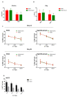
Function of expanded natural killer (eNK) cells using K562 and K562-HLA-E cells. Surface expression of CD107a (A) and intracellular expression of IFN-γ (B) of eNK cells were measured by incubation of eNK cells with K562 cells at an effector-to-target (E:T) ratio of 1:1 for 5 h followed by evaluation using flow cytometry. Bar graphs show the percentage of eNK cells for degranulation (CD107a) and IFN-γ (error bars, mean ± SD). (C) The cytotoxicity of eNK cells on days 14 and 49 toward K562 cells, and ADCC of eNK cells against rituximab-coated Raji cells were measured by carboxyfluorescein succinimidyl ester (CFSE)-based flow cytometry assay at E:T ratios of 1:1, 0.5:1, and 0.25:1 for 4 h. Results presented are the mean of 6 donors and error bars represent the mean ± SD. (D) The cytotoxicity of K562-HLA-E based eNK cells of C1C1 and C2C2 donors against MC7 (C2C2) target cells.

References

    1. Lanier L.L. Up on the tightrope: Natural killer cell activation and inhibition. Nat. Immunol. 2008;9:495–502. doi: 10.1038/ni1581. - DOI - PMC - PubMed
    1. Yokoyama W.M., Kim S., French A.R. The dynamic life of natural killer cells. Annu. Rev. Immunol. 2004;22:405–429. doi: 10.1146/annurev.immunol.22.012703.104711. - DOI - PubMed
    1. Gumá M., Angulo A., Vilches C., Gómez-Lozano N., Malats N., López-Botet M. Imprint of human cytomegalovirus infection on the NK cell receptor repertoire. Blood. 2004;104:3664–3671. doi: 10.1182/blood-2004-05-2058. - DOI - PubMed
    1. López-Botet M., Muntasell A., Vilches C. The CD94/NKG2C+ NK-cell subset on the edge of innate and adaptive immunity to human cytomegalovirus infection. Semin. Immunol. 2014;26:145–151. doi: 10.1016/j.smim.2014.03.002. - DOI - PubMed
    1. Cerwenka A., Lanier L.L. Natural killer cell memory in infection, inflammation and cancer. Nat. Rev. Immunol. 2016;16:112–123. doi: 10.1038/nri.2015.9. - DOI - PubMed

Substances