Review
. 2022 Aug 16;8(8):859.
doi: 10.3390/jof8080859.

Coccidioides Species: A Review of Basic Research: 2022

Affiliations
Review

Coccidioides Species: A Review of Basic Research: 2022

Theo N Kirkland et al. J Fungi (Basel). .

Abstract

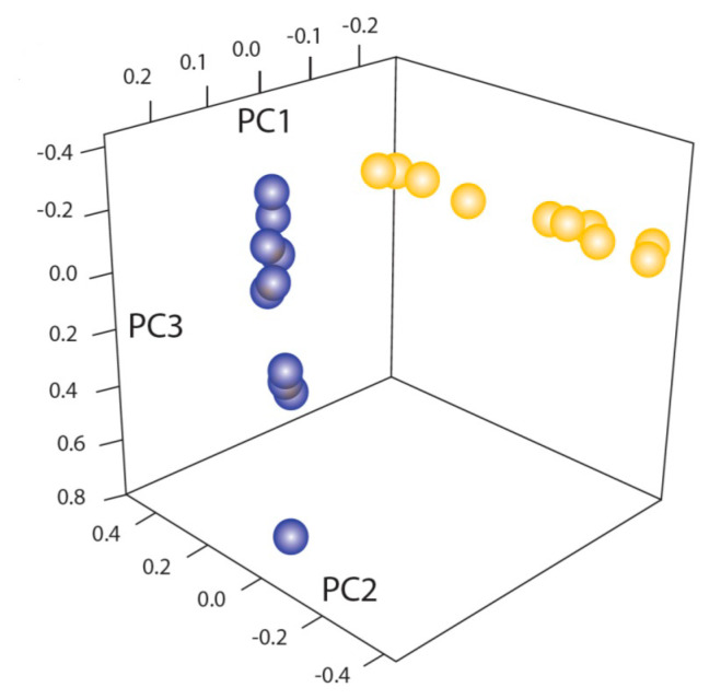
Coccidioides immitis and posadasii are closely related fungal species that cause coccidioidomycosis. These dimorphic organisms cause disease in immunocompetent as well as immunocompromised individuals and as much as 40% of the population is infected in the endemic area. Although most infections resolve spontaneously, the infection can be prolonged and, in some instances, fatal. Coccidioides has been studied for more than 100 years and many aspects of the organism and the disease it causes have been investigated. There are over 500 manuscripts concerning Coccidioides (excluding clinical articles) referenced in PubMed over the past 50 years, so there is a large body of evidence to review. We reviewed the most accurate and informative basic research studies of these fungi including some seminal older studies as well as an extensive review of current research. This is an attempt to gather the most important basic research studies about this fungus into one publication. To focus this review, we will discuss the mycology of the organism exclusively rather than the studies of the host response or clinical studies. We hope that this review will be a useful resource to those interested in Coccidioides and coccidioidomycosis.

Keywords: Coccidioides immitis; Coccidioides posadasii; coccidioidomycosis; dimorphic fungus; fungus; microbiology; mycelium; mycology; pathogenesis; spherule.


Conflict of interest statement

The authors report no conflict of interest. The funders played no role in the interpretation of data or the prepation of this manuscript.

Figures

Figure 1
Figure 1
Phylogenetic distance tree (Bayesian) with posterior probabilities based on 100 randomly selected single-copy orthologs (A). Phylogenetic categories used for gene family expansion/contraction and ortholog group analysis (B). N, newly sequenced genomes. C, Coccidioides, U, U. reesii; D, dermatophytes; Y, yeast-forming dimorphic fungal pathogens; O, outgroups. The figure is from [14].
Figure 2
Figure 2
Geographic distribution of CA and non-CA strains. Biogeographic range expansion into South America by C. immitis mirrors New World patterns of human migration. The colors refer to the genetic clusters on the left. * means: The asterisk marks an isolate from a patient who was diagnosed in Texas but had acquired the disease in California. The figure is from [21].
Figure 3
Figure 3
Temperature impacts growth ability of C. immitis isolates compared to C. posadasii on yeast extract media. Radial growth measurements at 37 °C for 46 C. posadasii and 39 C. immitis isolates in triplicate. The figure is from [23].
Figure 4
Figure 4
Updated map from the CDC estimating the distribution of Coccidioides spp. in the United States. This figure is from the Centers for Disease Control and Prevention (https://www.cdc.gov/fungal/diseases/coccidioidomycosis/causes.html) (accessed on 10 August 2022).
Figure 5
Figure 5
Arthroconidia within mycelia. Every alternate cell has degenerated. The bar is 5 μM. The figure is from the Centers for Disease Control and Prevention. (https://phil.cdc.gov/Details.aspx?pid=15780) (accessed on 10 August 2022).
Figure 6
Figure 6
Components of arthroconidia cell wall. A and B: Thin sections of conidia of C. immitis showing differentiation of wall layers. OW, outer wall layer; SW, septal walls of conidium; ICW, newly formed inner conidial wall composed of the outer zone (OZ) and more homogeneous inner zone (IZ); RL, rodlet layer; Pm, plasmalemma. Arrow locates soluble conidial wall fraction (SCWF) trapped between OW and RL which is released during the cell-shearing process. (A), magnification ×12,000; (B), magnification ×27,000. The figure is from [64] and is reproduced with permission. (C), composition of inner and outer cell wall and SCWF. The data for this figure is from [64].
Figure 7
Figure 7
The saprobic (mycelia and arthroconidia) and parasitic (spherule and endospore) life cycle. The figure is from [65]. (A), mycelia growing in the soil and differentiating into spherules in animals. (B), Endozoan-based life cycle of Coccidioides species. Beginning at the asterisk (*), arthroconidia travel from the hyphae that produced them short distances among small mammals in burrows, or longer distances above ground, to infect other animals. The arthroconidia convert to spherules and are either controlled by the immune reaction or develop endospores, which disseminate to produce grave disease. The infected animal dies, either from disseminated coccidioidomycosis or from other causes and, in either case, living Coccidioides present in the animal, now freed from the host immune system and living at lower temperatures, convert to hyphae. The hyphae grow through the dead animal and then produce abundant arthroconidia, which initiate a new cycle of life for the fungus. The figure is from [55] and is reproduced with permission.
Figure 7
Figure 7
The saprobic (mycelia and arthroconidia) and parasitic (spherule and endospore) life cycle. The figure is from [65]. (A), mycelia growing in the soil and differentiating into spherules in animals. (B), Endozoan-based life cycle of Coccidioides species. Beginning at the asterisk (*), arthroconidia travel from the hyphae that produced them short distances among small mammals in burrows, or longer distances above ground, to infect other animals. The arthroconidia convert to spherules and are either controlled by the immune reaction or develop endospores, which disseminate to produce grave disease. The infected animal dies, either from disseminated coccidioidomycosis or from other causes and, in either case, living Coccidioides present in the animal, now freed from the host immune system and living at lower temperatures, convert to hyphae. The hyphae grow through the dead animal and then produce abundant arthroconidia, which initiate a new cycle of life for the fungus. The figure is from [55] and is reproduced with permission.
Figure 8
Figure 8
Two-dimensional immuno-electrophoresis precipitin lines. Drawing of some typical precipitant lines in two-dimensional immuno-electrophoresis of coccidioidin with anti-coccidioidin antisera. The antigens are numbered as described [89].
Figure 9
Figure 9
Principal component analysis of the genomic sequence of C. immitis and C. posadasii strains. The blue points represent DNA sequence of C. immitis strains; the yellow points represent DNA sequence of C. posadasii strains. The figure is from [120].
Figure 10
Figure 10
Images in triplicate of spherules (A) labeled with calcofluor white (CFW) and FM4-64FX, a lipophilic dye (red) in vitro. SOW lipids are enriched in the inner leaflet of SOW. When the lipid is exposed as the SOW separates from the surface it binds to FM4-64FX dye [135]. Electron microscopy image (B) of a histological section of C. posadasii-infected tissue from the mouse lungs. Tissue section was labeled with osmium tetroxide (OsO4) to enhance the visibility of lipids. Host phagocytes are outlined with red dash-dot lines and their nuclei are labeled (N). Lipid-rich SOW fragments are visible inside phagocytes (white arrows). The bar is 5 μM. Image courtesy of Dr. Garry Cole and Dr. Kalpathi R. Seshan.
Figure 11
Figure 11
Comparison of differentially regulated genes in C. immitis and C. posadasii. Venn diagrams showing the number of genes commonly differentially regulated in the saprobic vs. parasitic growth phases of C. immitis and C. posadasii. The figure is from [15].
Figure 12
Figure 12
Biological GO terms enriched in spherules. Principle component analysis (PCA) plot of enriched GO terms of genes up-regulated in spherules, made using Revigo. The data for this figure is from [16].
Figure 13
Figure 13
(A). Wildtype and Δryp1 C. posadasii grown in Converse medium in spherule conditions. Spherules form in the WT organism; only hyphae with polar swelling but no spherules are seen in the Δryp1 C. posadasii mutant. The bars represent 10 μM. (B). Gene expression in the Δryp1 mutant. Morphology-dependent genes; genes differentially expressed in spherules compared to mycelia Theses figures are from [153].
Figure 14
Figure 14
Gene ontology enrichment analysis of the 27 biomarker candidates indicates enriched hydrolase activity and increased purine and carbohydrate metabolism functions. (A) Scatterplot of gene ontology enrichment of molecular functions, with size of the circles is proportional to the significance of enrichment. (B) Word cloud of enriched KEGG metabolic pathways. Size of the words indicates the significance of enrichment. Figures were produced using Revigo and GOSummaries. The figure is from [77,78].

References

    1. CDC Increase in reported coccidioidomycosis—United States, 1998–2011. Morb. Mortal. Wkly. Rep. 2013;62:217–221. - PMC - PubMed
    1. Hector R.F., Rutherford G.W., Tsang C.A., Erhart L.M., McCotter O., Anderson S.M., Komatsu K., Tabnak F., Vugia D.J., Yang Y., et al. The public health impact of coccidioidomycosis in Arizona and California. Int. J. Environ. Res. Public Health. 2011;8:1150–1173. doi: 10.3390/ijerph8041150. - DOI - PMC - PubMed
    1. Valdivia L., Nix D., Wright M., Lindberg E., Fagan T., Lieberman D., Stoffer T.P., Ampel N.M., Galgiani J.N. Coccidioidomycosis as a common cause of community-acquired pneumonia. Emerg. Inf. Dis. 2006;12:958–962. doi: 10.3201/eid1206.060028. - DOI - PMC - PubMed
    1. Grizzle A.J., Wilson L., Nix D.E., Galgiani J.N. Clinical and Economic Burden of Valley Fever in Arizona: An Incidence-Based Cost-of-Illness Analysis. Open Forum Infect. Dis. 2021;8:ofaa623. doi: 10.1093/ofid/ofaa623. - DOI - PMC - PubMed
    1. Deresinski S., Mirels L.F. Coccidioidomycosis: What a long strange trip it’s been. Med. Mycol. 2019;57:S3–S15. doi: 10.1093/mmy/myy123. - DOI - PMC - PubMed