Review
. 2022 Aug 14;11(16):4742.
doi: 10.3390/jcm11164742.

A Study on the Pathogenesis of Vascular Cognitive Impairment and Dementia: The Chronic Cerebral Hypoperfusion Hypothesis

Affiliations
Review

A Study on the Pathogenesis of Vascular Cognitive Impairment and Dementia: The Chronic Cerebral Hypoperfusion Hypothesis

Weiwei Yu et al. J Clin Med. .

Abstract

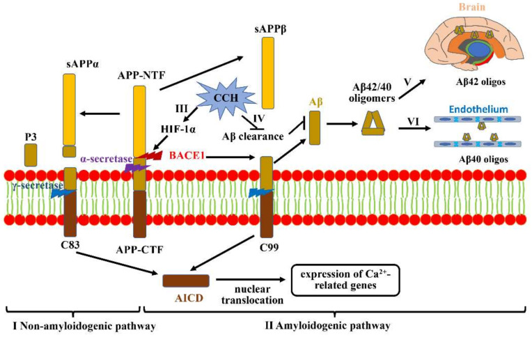
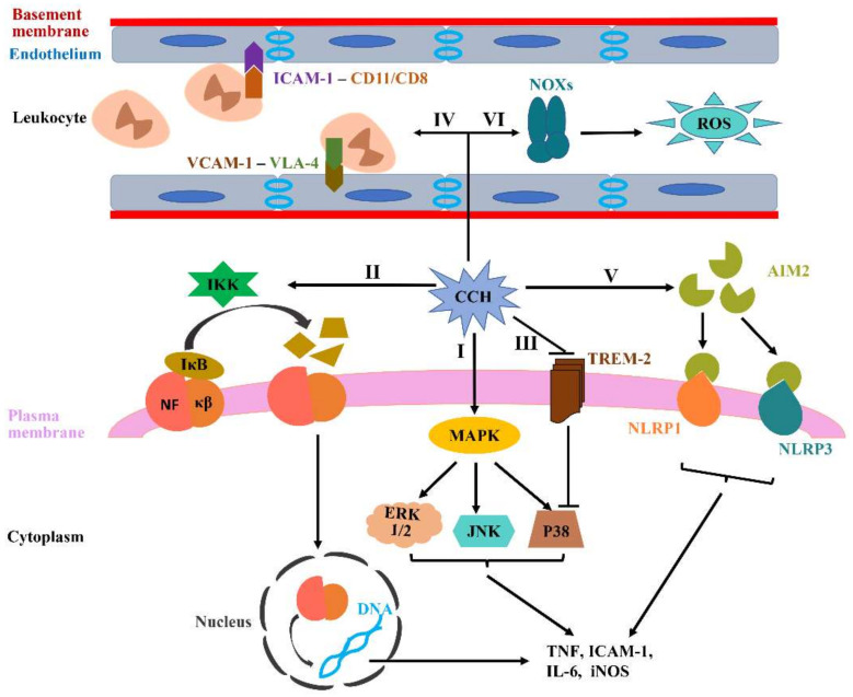
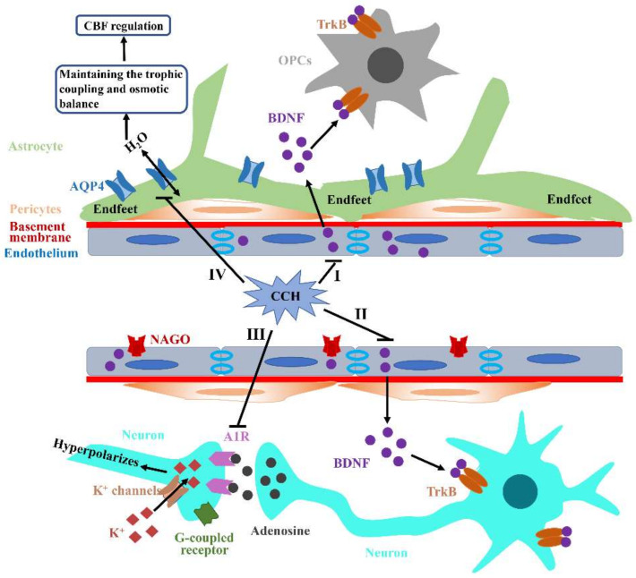
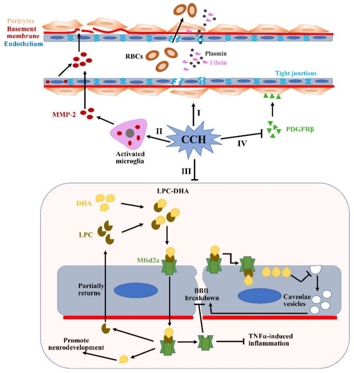
The pathogenic mechanisms underlying vascular cognitive impairment and dementia (VCID) remain controversial due to the heterogeneity of vascular causes and complexity of disease neuropathology. However, one common feature shared among all these vascular causes is cerebral blood flow (CBF) dysregulation, and chronic cerebral hypoperfusion (CCH) is the universal consequence of CBF dysregulation, which subsequently results in an insufficient blood supply to the brain, ultimately contributing to VCID. The purpose of this comprehensive review is to emphasize the important contributions of CCH to VCID and illustrate the current findings about the mechanisms involved in CCH-induced VCID pathological changes. Specifically, evidence is mainly provided to support the molecular mechanisms, including Aβ accumulation, inflammation, oxidative stress, blood-brain barrier (BBB) disruption, trophic uncoupling and white matter lesions (WMLs). Notably, there are close interactions among these multiple mechanisms, and further research is necessary to elucidate the hitherto unsolved questions regarding these interactions. An enhanced understanding of the pathological features in preclinical models could provide a theoretical basis, ultimately achieving the shift from treatment to prevention.

Keywords: blood-brain barrier; cerebral blood flow regulation; chronic cerebral hypoperfusion; neuroinflammation; oxidative stress; trophic uncoupling; vascular cognitive impairment and dementia; white matter lesions; β-amyloid.


Conflict of interest statement

The authors declare no conflict of interest.

Figures

Figure 1
Figure 1
Aβ accumulation and aggravation under CCH. (I) Non-amyloidogenic pathway: APP is first cleaved by α-secretase to generate sAPPα and C83. C83 then produces P3 and AICD under the action of γ-secretase, which is shifted to the nucleus to regulate Ca2+-related gene expression. (II) Amyloidogenic pathway: full-length APP can also be cleaved by BACE1 to release soluble APPβ from the cell membrane and retain C99. Subsequent cleavage of C99 by γ-secretase generates Aβ42/40. (III) CCH-mediated hypoxia can promote BACE1 gene expression by upregulating HIF-1α. (IV) CCH is also likely to increase Aβ accumulation via impairing Aβ clearance. (V) The more aggressive Aβ42 tends to deposit in vulnerable brain regions that are primarily associated with learning and memory. (VI) Aβ40 prefers to deposit in non-neuronal cells, mainly including cerebral vessels.
Figure 2
Figure 2
CCH-induced neuroinflammation and oxidative stress. (IIII) The activated microglia by CCH can generate various pro-inflammatory cytokines, such as IL-1β, IL-6 and TNF, in which multiple signaling pathways are involved, including MAPKs, NF-κβ and TREM-2. (IV) CCH can directly accelerate endothelium activation. Activated endothelium express more ICAM-1 and VCAM-1 to bind to CD11/CD18 and VLA-4, which contribute to the adherence and extravasation of leukocytes into brain parenchyma. (V) AIM2 inflammasome signaling can be activated during CCH, which increases microglial activation and subsequently promotes the generation of pro-inflammatory cytokines. (VI) NOXs, recognized as the primary source of ROS in the brain, are thought to be the key contributing factors to CCH-related oxidative stress.
Figure 3
Figure 3
Aβ accumulation and aggravation under CCH. (I,II) The abundant BDNF produced by endothelium in the NVU can bind to TrkB receptors on OPCs and neurons to promote their proliferation, migration and survival, which can be disrupted by CCH. (III) CCH can significantly decrease A1R expression and subsequently impede neuron hyperpolarizes in the post-synapse through G-protein coupled inwardly rectifying K+ channels. (IV) AQP4, as a water channel, is generally restricted to astrocytic end-feet processes that mainly contact synapses and vasculature. Additionally, AQP4 also plays an important role in maintaining osmotic balance through eliminating edema induced by various damages. CCH can lead to AQP4 mislocalization and then induce extensive trophic uncoupling and osmotic imbalance, ultimately destroying CBF regulation and attenuating perfusion to the brain.
Figure 4
Figure 4
Potential mechanisms of BBB breakdown during CCH. (I) Pericytes detach from the perivascular positions under the CCH condition, which subsequently reduce the expression of tight junction proteins. Additionally, CCH can also directly disrupt the expressions of tight junction proteins. (II) CCH can upregulate levels of MMP2 expression in the activated microglia and endothelium. Increased MMP2 can accelerate the degradation of many major ECM constituents, such as collagen IV, fibronectin and gelatin. (III) Mfsd2a plays a critical role in transporting DHA into the cells and maintaining the low rate of vesicular transcytosis. However, the expression of the Mfsd2a protein is significantly reduced during CCH, which causes augmented vesicular transcytosis. (IV) CCH-mediated PDGFRβ reduction can result in significantly decreased microvessel lengths. All of these pathological changes induced by CCH can subsequently lead to significant BBB impairment and contribute to the translation of many substances from the blood vessels to the brain parenchyma, such as RBCs, plasmin and fibrin. RBCs: red blood cells; DHA: docosahexaenoic acid; LPC: lysophosphatidylcholine.

References

    1. Jellinger K.A. Pathology and pathogenesis of vascular cognitive impairment—A critical update. Front. Aging Neurosci. 2013;5:17. doi: 10.3389/fnagi.2013.00017. - DOI - PMC - PubMed
    1. Iadecola C. The Pathobiology of Vascular Dementia. Neuron. 2013;80:844–866. doi: 10.1016/j.neuron.2013.10.008. - DOI - PMC - PubMed
    1. Frantellizzi V., Pani A., Ricci M., Locuratolo N., Fattapposta F., De Vincentis G. Neuroimaging in Vascular Cognitive Impairment and Dementia: A Systematic Review. J. Alzheimers Dis. 2020;73:1279–1294. doi: 10.3233/JAD-191046. - DOI - PubMed
    1. Yang T., Sun Y., Lu Z., Leak R., Zhang F. The impact of cerebrovascular aging on vascular cognitive impairment and dementia. Ageing Res. Rev. 2016;34:15–29. doi: 10.1016/j.arr.2016.09.007. - DOI - PMC - PubMed
    1. Mijajlović M.D., Pavlović A., Brainin M., Heiss W.D., Quinn T.J., Ihle-Hansen H.B., Hermann D.M., Assayag E.B., Richard E., Thiel A., et al. Post-stroke dementia—A comprehensive review. BMC Med. 2017;15:11. doi: 10.1186/s12916-017-0779-7. - DOI - PMC - PubMed

LinkOut - more resources