Review
. 2022 Aug 17;11(16):4807.
doi: 10.3390/jcm11164807.

An Update on the Management of Acute High-Risk Pulmonary Embolism

Affiliations
Review

An Update on the Management of Acute High-Risk Pulmonary Embolism

Romain Chopard et al. J Clin Med. .

Abstract

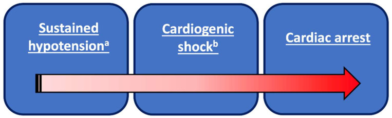
Hemodynamic instability and right ventricular (RV) dysfunction are the key determinants of short-term prognosis in patients with acute pulmonary embolism (PE). High-risk PE encompasses a wide spectrum of clinical situations from sustained hypotension to cardiac arrest. Early recognition and treatment tailored to each individual are crucial. Systemic fibrinolysis is the first-line pulmonary reperfusion therapy to rapidly reverse RV overload and hemodynamic collapse, at the cost of a significant rate of bleeding. Catheter-directed pharmacological and mechanical techniques ensure swift recovery of echocardiographic parameters and may possess a better safety profile than systemic thrombolysis. Further clinical studies are mandatory to clarify which pulmonary reperfusion strategy may improve early clinical outcomes and fill existing gaps in the evidence.

Keywords: catheter-based therapy; high-risk pulmonary embolism; multidisciplinary care team; surgical embolectomy; systemic thrombolysis.


Conflict of interest statement

The authors declare no conflict of interest.

Figures

Figure 1
Figure 1
Clinical spectrum of high-risk pulmonary embolism. (a) Systolic blood pressure (BP) < 90 mmHg or systolic BP drop ≥ 40 mmHg, lasting longer than 15 min, and not caused by new-onset arrhythmia, hypovolemia, or sepsis; (b) systolic BP < 90 mmHg or vasopressors required to achieve a BP ≥ 90 mmHg despite adequate filling status and end-organ hypoperfusion (altered mental status; cold, clammy skin; oliguria/anuria; increased serum lactate > 2.4 mmol/L).
Figure 2
Figure 2
Trans-thoracic parameters for the assessment of right ventricular dysfunction in the acute phase of pulmonary embolism. (A). Enlarged right ventricle (parasternal long axis view). (B). Flattened intraventricular septum (arrows) (parasternal view). (C). Mobile thrombus (arrows) in the right heart cavities. (D). Decreased peak systolic (S’) velocity of tricuspid annulus < 9.5 cm/s (Tissue Doppler imaging). (E). Dilated right ventricle with basal RV/LV ratio > 1.0 (double-ended arrows), and McConnell sign (i.e., akinesia of the mid free wall (arrows) with normal motion at the apex hypokinesia of the RV) (four chamber view). (F). 60/60 sign: Association of acceleration time of pulmonary ejection < 60 ms and midsystolic “notch” with mildly elevated (<60 mmHg) peak systolic gradient at the tricuspid valve. (G). Decreased tricuspid annular plane systolic excursion (TAPSE) < 16 mm (M-Mode). (H). Distended inferior vena cava with diminished inspiratory collapsibility (subcostal view). RV, right ventricle; LV, left ventricle; Ao, aorta; RA, right atrium; LA, left atrium.
Figure 3
Figure 3
Direct vascular signs (A), indirect vascular signs (B), and parenchymal changes (C) on computed tomography scan between acute pulmonary embolism and chronic thrombo-embolism pulmonary hypertension (CTEPH).
Figure 3
Figure 3
Direct vascular signs (A), indirect vascular signs (B), and parenchymal changes (C) on computed tomography scan between acute pulmonary embolism and chronic thrombo-embolism pulmonary hypertension (CTEPH).
Figure 3
Figure 3
Direct vascular signs (A), indirect vascular signs (B), and parenchymal changes (C) on computed tomography scan between acute pulmonary embolism and chronic thrombo-embolism pulmonary hypertension (CTEPH).

Similar articles

Cited by

References

    1. Wendelboe A.M., Raskob G.E. Global Burden of Thrombosis: Epidemiologic Aspects. Circ. Res. 2016;118:1340–1347. doi: 10.1161/CIRCRESAHA.115.306841. - DOI - PubMed
    1. Goldhaber S.Z., Bounameaux H. Pulmonary embolism and deep vein thrombosis. Lancet. 2012;379:1835–1846. doi: 10.1016/S0140-6736(11)61904-1. - DOI - PubMed
    1. Konstantinides S.V., Meyer G., Becattini C., Bueno H., Geersing G.J., Harjola V.P., Huisman M.V., Humbert M., Jennings C.S., Jimenez D., et al. 2019 ESC Guidelines for the diagnosis and management of acute pulmonary embolism developed in collaboration with the European Respiratory Society (ERS) Eur. Heart J. 2020;41:543–603. doi: 10.1093/eurheartj/ehz405. - DOI - PubMed
    1. Stevens S.M., Woller S.C., Kreuziger L.B., Bounameaux H., Doerschug K., Geersing G.J., Huisman M.V., Kearon C., King C.S., Knighton A.J., et al. Antithrombotic Therapy for VTE Disease: Second Update of the CHEST Guideline and Expert Panel Report. Chest. 2021;160:e545–e608. doi: 10.1016/j.chest.2021.07.055. - DOI - PubMed
    1. Becattini C., Agnelli G., Lankeit M., Masotti L., Pruszczyk P., Casazza F., Vanni S., Nitti C., Kamphuisen P., Vedovati M.C., et al. Acute pulmonary embolism: Mortality prediction by the 2014 European Society of Cardiology risk stratification model. Eur. Respir. J. 2016;48:780–786. doi: 10.1183/13993003.00024-2016. - DOI - PubMed

LinkOut - more resources