Review
. 2022 Aug 20;11(16):4896.
doi: 10.3390/jcm11164896.

COVID-19-Related ARDS: Key Mechanistic Features and Treatments

Affiliations
Review

COVID-19-Related ARDS: Key Mechanistic Features and Treatments

John Selickman et al. J Clin Med. .

Abstract

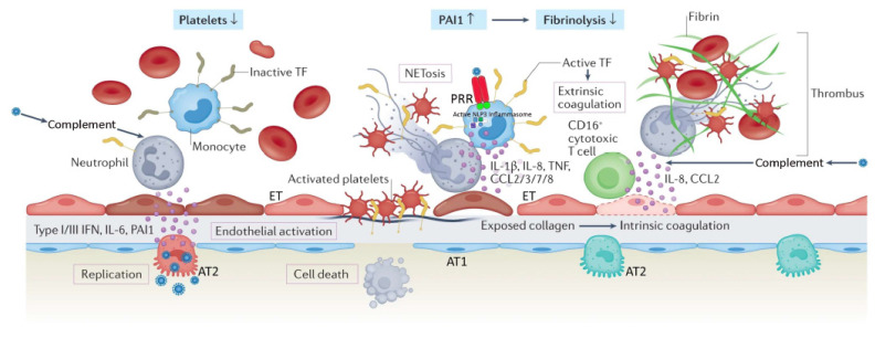
Acute respiratory distress syndrome (ARDS) is a heterogeneous syndrome historically characterized by the presence of severe hypoxemia, high-permeability pulmonary edema manifesting as diffuse alveolar infiltrate on chest radiograph, and reduced compliance of the integrated respiratory system as a result of widespread compressive atelectasis and fluid-filled alveoli. Coronavirus disease 19 (COVID-19)-associated ARDS (C-ARDS) is a novel etiology caused by severe acute respiratory syndrome coronavirus 2 (SARS-CoV-2) that may present with distinct clinical features as a result of the viral pathobiology unique to SARS-CoV-2. In particular, severe injury to the pulmonary vascular endothelium, accompanied by the presence of diffuse microthrombi in the pulmonary microcirculation, can lead to a clinical presentation in which the severity of impaired gas exchange becomes uncoupled from lung capacity and respiratory mechanics. The purpose of this review is to highlight the key mechanistic features of C-ARDS and to discuss the implications these features have on its treatment. In some patients with C-ARDS, rigid adherence to guidelines derived from clinical trials in the pre-COVID era may not be appropriate.

Keywords: COVID-19; SARS-CoV-2; acute respiratory distress syndrome; mechanical ventilation.


Conflict of interest statement

The authors declare no conflict of interest.

Figures

Figure 1
Figure 1
Severe coronavirus disease 19 is characterized by immune cell-mediated hypercoagulability and hypofibrinolysis. Hypoxia, cytokines, chemokines, damage-associated molecular patterns, and direct infection by the virus contribute to alveolar and endothelial cell death, and disruption of the alveolar–capillary barrier. Exposed extracellular matrix can trigger both the extrinsic coagulation (via tissue factor (TF)) and the intrinsic coagulation (via collagen/RNA). Recruited monocytes (with virus-activated NLP3 inflammasomes) and neutrophils amplify the inflammatory response, as well as the activation of coagulation by expressing active tissue factor (TF) and releasing neutrophil extracellular traps (NETs), respectively. Complement activation by the virus promotes active TF expression by neutrophils, and differentiation of cytotoxic CD-16+ T cells. NETs recruit platelets, which are subsequently activated by NET histones and the C3a and C5a complement fragments; this results in platelet release of cytokines. Activated platelets secrete coagulation-sustaining factors. The immunothrombotic process leads to diffuse small-vessel thromboses and thrombocytopenia. Concurrently, increased expression of plasminogen activator inhibitor (PA1) attenuates fibrinolysis. AT1, alveolar type 1 cell; AT2, alveolar type 2 cell; ET, endothelial cell; PRR, pattern recognition receptor; IL, interleukin; CCL, CC chemokine ligand; IFN, interferon. Reproduced in part with permission from [26]; copyright (2022) by Springer Nature.
Figure 2
Figure 2
Diagrammatic presentation of physiological mechanisms associated with pronation in acute respiratory distress syndrome (ARDS). (A,C) show the shape of lung units (i.e., alveoli) without the effect of gravity. (B) In the supine position, the volume of dorsal lung units is significantly smaller than the volume of ventral lung units, as a result of gravity and pleural pressure; thus, ventral lung units are more prone to overdistention and dorsal lung units are more prone to compression atelectasis. (D) In the prone position, gravity and pleural pressure result in a decrease in the volume of the ventral lung units and an increase in the volume of the dorsal lung units. (E) In the supine position, the ventral transpulmonary pressure (PTP) may substantially exceed the dorsal PTP (F) Prone positioning reduces the ventral-to-dorsal PTP gradient thereby augmenting the homogeneity of ventilation. (G) The reopening, dorsal lung units continue to receive most of the blood flow. (H) The ventral lung units may exhibit a greater tendency to collapse, but are still relatively underperfused. Reproduced in concordance with the Creative Commons Attribution License (CC-BY) from [138].

Similar articles

Cited by

References

    1. Ashbaugh D.G., Bigelow D.B., Petty T.L., Levine B.E. Acute respiratory distress in adults. Lancet. 1967;2:319–323. doi: 10.1016/S0140-6736(67)90168-7. - DOI - PubMed
    1. Gattinoni L., Marini J.J. Isn’t it time to abandon ARDS? The COVID-19 lesson. Crit. Care. 2021;25:326. doi: 10.1186/s13054-021-03748-6. - DOI - PMC - PubMed
    1. Bernard G.R. Acute respiratory distress syndrome: A historical perspective. Am. J. Respir. Crit. Care Med. 2005;172:798–806. doi: 10.1164/rccm.200504-663OE. - DOI - PMC - PubMed
    1. Thille A.W., Esteban A., Fernandez-Segoviano P., Rodriguez J.M., Aramburu J.A., Penuelas O., Cortes-Puch I., Cardinal-Fernandez P., Lorente J.A., Frutos-Vivar F. Comparison of the Berlin definition for acute respiratory distress syndrome with autopsy. Am. J. Respir. Crit. Care Med. 2013;187:761–767. doi: 10.1164/rccm.201211-1981OC. - DOI - PubMed
    1. Marini J.J. Limitations of clinical trials in acute lung injury and acute respiratory distress syndrome. Curr. Opin. Crit. Care. 2006;12:25–31. doi: 10.1097/01.ccx.0000198996.22072.4a. - DOI - PubMed

LinkOut - more resources