Review
. 2022 Aug 10;12(8):1215.
doi: 10.3390/life12081215.

Imaging of Cerebral Venous Thrombosis

Affiliations
Review

Imaging of Cerebral Venous Thrombosis

Jean-Claude Sadik et al. Life (Basel). .

Abstract

Cerebral venous thrombosis is a rare cause of stroke. Imaging is essential for diagnosis. Although digital subtraction angiography is still considered by many to be the gold standard, it no longer plays a significant role in the diagnosis of cerebral venous thrombosis. MRI, which allows for imaging the parenchyma, vessels and clots, and CT are the reference techniques. CT is useful in case of contraindication to MRI. After presenting the radio-anatomy for MRI, we present the different MRI and CT acquisitions, their pitfalls and their limitations in the diagnosis of cerebral venous thrombosis.

Keywords: CT venography; MR venography; MRI; cerebral vein; cerebral venous thrombosis; dural sinus thrombosis; intracranial hypertension; venous stroke.


Conflict of interest statement

Julien Savatovsky received honoraria for lectures and board participation from Bayer Healthcare. The other authors declare no conflict of interest.

Figures

Figure 1
Figure 1
Sagittal fluid attenuated inversion recovery (FLAIR) image. Spontaneous subarachnoid hemorrhage in the precentral, central, and postcentral sulci in the context of cerebral venous thrombosis. Some cortical veins are visible, but their thrombosis cannot be verified because they are drowned in the spontaneous FLAIR signal of the subarachnoid hemorrhage.
Figure 2
Figure 2
Digital subtraction angiography, sagittal plane. Green arrow: superior sagittal sinus. Yellow arrow: inferior sagittal sinus. Blue arrow: straight sinus. Orange arrow: lateral sinus. Red arrow: torcular Herophili.
Figure 3
Figure 3
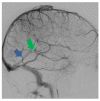
Digital subtraction angiography, sagittal plane. Red arrow: superficial middle cerebral vein. Green arrow: superior anastomotic vein (Trolard’s vein). Blue arrow: inferior anastomotic vein (Labbé’s vein).
Figure 4
Figure 4
Digital subtraction angiography, sagittal plane. Anatomy of Galen’s ampulla and straight sinus. The straight sinus receives blood from the inferior sagittal sinus and the vein of Galen. It flows into the torcular Herophili, where it joins the superior sagittal sinus. Green arrow: Galen’s ampulla. Blue arrow: straight sinus.
Figure 5
Figure 5
CE-MRV, axial and sagittal planes. MR anatomy of the basal veins. Red arrow: basal veins (former basal veins of Rosenthal).
Figure 6
Figure 6
CE-MRV, axial and sagittal planes. MR anatomy of the septal, thalamostriate, and internal cerebral veins. Blue arrow: septal veins. Green arrow: thalamostriate veins. Yellow arrow: internal cerebral veins.
Figure 7
Figure 7
Decisional algorithm for choosing arterial or venous MR angiography. PC: phase contrast, Gd, gadolinium, TOF, time of flight.
Figure 8
Figure 8
MRI sequences for imaging the veins, the thrombus, the parenchyma, and the meninges [15]. PC: phase contrast, TOF: time of flight, ASL: arterial spin labeling, CTA: CT angiography. 3D CE-MRA (ATECO): 3D contrast-enhanced MR angiography (auto-triggered elliptic centric ordered), 4D (TRAK, TRICKS, TWIST): dynamic contrast-enhanced MR angiography, FLAIR: fluid-attenuated inversion recovery, Diffusion: diffusion-weighted imaging, SWI: susceptibility-weighted imaging.
Figure 9
Figure 9
(a) Nonenhanced T1-weighted images, sagittal plane. Spontaneous hypersignal of the superior sagittal sinus suggests thrombosis. (b) T2* images, axial plane. Spontaneous hyposignal of the cortical veins of the vertex suggests cortical vein thrombi.
Figure 10
Figure 10
From left to right, spontaneous signal of the right lateral sinus suggesting thrombosis: (a) T1 hypersignal, (b) T2* hyposignal, (c) FLAIR hypersignal, (d) diffusion-weighted imaging hypersignal.
Figure 11
Figure 11
3D contrast-enhanced T1-weighted gradient-echo images. (a) Sagittal plane; “empty delta” sign in the lateral sinus. (b) Axial plane; empty delta sign in the superior sagittal sinus. The enhancement related to the injection occurs around the thrombus, which appears in relative hyposignal.
Figure 12
Figure 12
CE-MRV. From left to right: (a,b) maximum intensity projection (MIP) reconstructions; interruption of the proximal portion of the right lateral sinus; (c,d) native slices; defect in the lateral sinus suggests a thrombus.
Figure 13
Figure 13
MRI signs of intracranial hypertension. They illustrate common findings in intracranial hypertension regardless of the presence of cerebral venous thrombosis. From left to right: (a) optic disc FLAIR hyperintensity, (b) dilation of the optic nerve sheaths, (c) empty sella turcica.
Figure 14
Figure 14
From left to right: (a) contrast-enhanced T1 spin echo, (b) contrast-enhanced T1 gradient echo.
Figure 15
Figure 15
Nonenhanced CT. Spontaneous hyperdensity of the thrombus in the right lateral/sigmoid sinus junction.
Figure 16
Figure 16
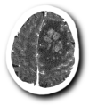
Left frontopolar venous hemorrhagic softening on CT.
Figure 17
Figure 17
CT. From left to right: (a) nonenhanced CT: spontaneous hyperdensity of the superior sagittal sinus, (b) enhanced CT: empty delta sign testifying to the circulation of contrast around the thrombus, which appears as a defect of signal in the center of the superior sagittal sinus.
Figure 18
Figure 18
Volume rendering reconstructions of cerebral CT venography. Thrombosis of the anterior part of the superior sagittal sinus.
Figure 19
Figure 19
From left to right: (a) nonenhanced CT, sagittal plane; (spontaneous hyperdensity of the straight sinus); (b) after the injection of iodinated contrast media, absence of opacification of the straight sinus, thrombosis of the superior sagittal sinus above the torcular Herophili; (c) MRI, nonenhanced sagittal T1 sequence: spontaneous hypersignal of the thrombus.
Figure 20
Figure 20
From left to right: (a,b) susceptibility-weighted imaging, axial (a) and sagittal (b) planes (thrombosis of the internal cerebral and basilar veins and straight sinus); (c) T1 sequence sagittal plane (spontaneous hypersignal of the thrombus in the straight sinus); (d) FLAIR image, axial plane (spontaneous hypersignal of the thrombus in the left lateral sinus).
Figure 21
Figure 21
Ischemic lesion of the right thalamus. (a) FLAIR image, axial plane (spontaneous hypersignal of the right thalamus); (b) diffusion-weighted imaging; (hypersignal of the right thalamus); (c) nonenhanced T1 image, sagittal plane; (hyposignal of the right thalamus); (d) nonenhanced CT image (hypodensity of the right thalamus); (e) thrombosis of the right internal cerebral vein. Note the presence of left frontal sinusitis.
Figure 21
Figure 21
Ischemic lesion of the right thalamus. (a) FLAIR image, axial plane (spontaneous hypersignal of the right thalamus); (b) diffusion-weighted imaging; (hypersignal of the right thalamus); (c) nonenhanced T1 image, sagittal plane; (hyposignal of the right thalamus); (d) nonenhanced CT image (hypodensity of the right thalamus); (e) thrombosis of the right internal cerebral vein. Note the presence of left frontal sinusitis.
Figure 22
Figure 22
Left lateral sinus hypoplasia: (a) sagittal plane of the right lateral sinus; (b) phase contrast, lack of filling of the left lateral sinus; (c) sagittal plane of the right lateral sinus confirming its small caliber.
Figure 23
Figure 23
(a) Contrast-enhanced T1 image, sagittal plane (filling defect in the left lateral sinus); (b) CE-MRV confirms the typical round aspect of Pacchionian arachnoid granulations.

References

    1. Ulivi L., Squitieri M., Cohen H., Cowley P., Werring D.J. Cerebral venous thrombosis: A practical guide. Pract. Neurol. 2020;20:356–367. doi: 10.1136/practneurol-2019-002415. - DOI - PubMed
    1. Jianu D.C., Jianu S.N., Munteanu G., Flavius T.D., Brasan C. Ischemic Stroke of Brain. Volume 1. IntechOpen; London, UK: 2018. pp. 45–76.
    1. Stam J. Thrombosis of the cerebral veins and sinuses. N. Engl. J. Med. 2005;352:1791–1798. doi: 10.1056/NEJMra042354. - DOI - PubMed
    1. Bousser M.-G., Ferro J.M. Cerebral venous thrombosis: An update. Lancet Neurol. 2007;6:162–170. doi: 10.1016/S1474-4422(07)70029-7. - DOI - PubMed
    1. Ferro J.M., Bousser M., Canhão P., Coutinho J.M., Crassard I., Dentali F., di Minno M., Maino A., Martinelli I., Masuhr F., et al. European Stroke Organization guideline for the diagnosis and treatment of cerebral venous thrombosis—Endorsed by the European Academy of Neurology. Eur. J. Neurol. 2017;24:1203–1213. doi: 10.1111/ene.13381. - DOI - PubMed

LinkOut - more resources