

Comparison of Anatomical Conformity between TomoFix Anatomical Plate and TomoFix Conventional Plate in Open-Wedge High Tibial Osteotomy
- PMID: 36013511
- PMCID: PMC9413536
- DOI: 10.3390/medicina58081045
Comparison of Anatomical Conformity between TomoFix Anatomical Plate and TomoFix Conventional Plate in Open-Wedge High Tibial Osteotomy
Abstract
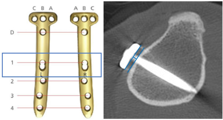
Background and Objectives: The TomoFix anatomical plate was developed to improve plate position, proximal screw direction, and post-correction tibial contouring. The purpose of this study was to compare postoperative configurations between the TomoFix anatomical plate and the TomoFix conventional plate. It was hypothesized that the new modified plate provides a better fixative coaptation than the conventional plate. Materials and Methods: A total of 116 cases (112 patients) were enrolled in this study from March 2015 to February 2021. Among them, 63 patients underwent surgery using the TomoFix conventional plate, and 53 underwent surgery using the TomoFix anatomical plate. The radiographic outcomes, including the hip−knee−ankle (HKA) angle, medial proximal tibial angle (MPTA), tibial slope, plate angle, proximal screw angles, and plate-to-cortex distance at #1 hole (just below the osteotomy site) were compared between the two groups. Results: Patients with the TomoFix anatomical plate showed similar results in terms of the pre- and postoperative HKA angle, MPTA, and tibial slope. The TomoFix anatomical group showed a significantly greater plate angle (39.2° ± 8.1° vs. 31.7° ± 7.0°, p < 0.001) and less screw angles, indicating that the TomoFix anatomical plates allowed a more posterior plate position than the conventional plate. The plate-to-cortex distance was significantly less in the TomoFix anatomical group than in the TomoFix conventional group (p < 0.001). Conclusion: The TomoFix anatomical plate showed a more posteromedial plating position, better proximal screw direction to the lateral hinge, and improved post-correction tibial contour compared to the TomoFix conventional plate.
Keywords: TomoFix; anatomical conformity; high tibial osteotomy; plate position.
Conflict of interest statement
The authors declare that they have no conflict of interest.
Figures


Similar articles
-
Biplanar Open Wedge High Tibial Osteotomy in the Medial Compartment Osteoarthritis of the Knee Joint: Comparison between the Aescula and TomoFix Plate.Clin Orthop Surg. 2015 Jun;7(2):185-90. doi: 10.4055/cios.2015.7.2.185. Epub 2015 May 18. Clin Orthop Surg. 2015. PMID: 26217464 Free PMC article.
-
Comparison of the clinical and radiological outcomes of Puddu and TomoFix plates for medial opening-wedge high tibial osteotomy: A two-year follow-up of a randomized controlled trial.Orthop Traumatol Surg Res. 2025 Feb;111(1):103845. doi: 10.1016/j.otsr.2024.103845. Epub 2024 Feb 23. Orthop Traumatol Surg Res. 2025. PMID: 38403264 Clinical Trial.
-
Plate removal without loss of correction after open-wedge high tibial osteotomy is possible when posterior cortex bone union reaches osteotomy gap center even in incompletely filled gaps.Knee Surg Sports Traumatol Arthrosc. 2020 Jun;28(6):1827-1834. doi: 10.1007/s00167-019-05615-y. Epub 2019 Jul 4. Knee Surg Sports Traumatol Arthrosc. 2020. PMID: 31273409
-
Patient-specific high tibial osteotomy for varus malalignment: 3D-printed plating technique and review of the literature.Eur J Orthop Surg Traumatol. 2022 Jul;32(5):845-855. doi: 10.1007/s00590-021-03043-8. Epub 2021 Jun 20. Eur J Orthop Surg Traumatol. 2022. PMID: 34148123 Review.
-
Accuracy of frontal and sagittal plane correction in open-wedge high tibial osteotomy.Arthroscopy. 2004 Apr;20(4):366-72. doi: 10.1016/j.arthro.2004.01.024. Arthroscopy. 2004. PMID: 15067275 Review.
Cited by
-
Risk Factors and Clinical Outcomes of Osteotomy Plane Violation by D-Hole Screws in Medial Open Wedge High Tibial Osteotomy: A Simulation and Comparative Study.Medicina (Kaunas). 2023 Nov 30;59(12):2104. doi: 10.3390/medicina59122104. Medicina (Kaunas). 2023. PMID: 38138208 Free PMC article. Clinical Trial.
-
Are TomoFix Locking Plates Really Anatomical for Indian Population?Indian J Orthop. 2024 Mar 15;58(5):495-502. doi: 10.1007/s43465-024-01119-1. eCollection 2024 May. Indian J Orthop. 2024. PMID: 38694700 Free PMC article.
References
MeSH terms
LinkOut - more resources
Full Text Sources
