. 2022 Aug 8;58(8):1070.
doi: 10.3390/medicina58081070.

Acute Posterior Multifocal Placoid Pigment Epitheliopathy (APMPPE): A Comprehensive Approach and Case Series: Systemic Corticosteroid Therapy Is Necessary in a Large Proportion of Cases

Affiliations

Acute Posterior Multifocal Placoid Pigment Epitheliopathy (APMPPE): A Comprehensive Approach and Case Series: Systemic Corticosteroid Therapy Is Necessary in a Large Proportion of Cases

Ioannis Papasavvas et al. Medicina (Kaunas). .

Abstract

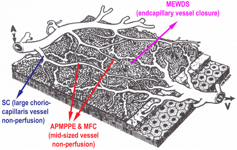
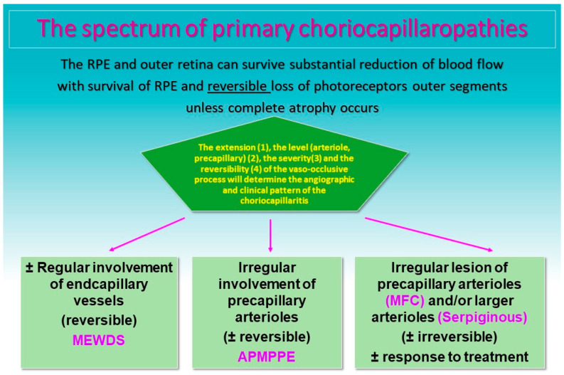
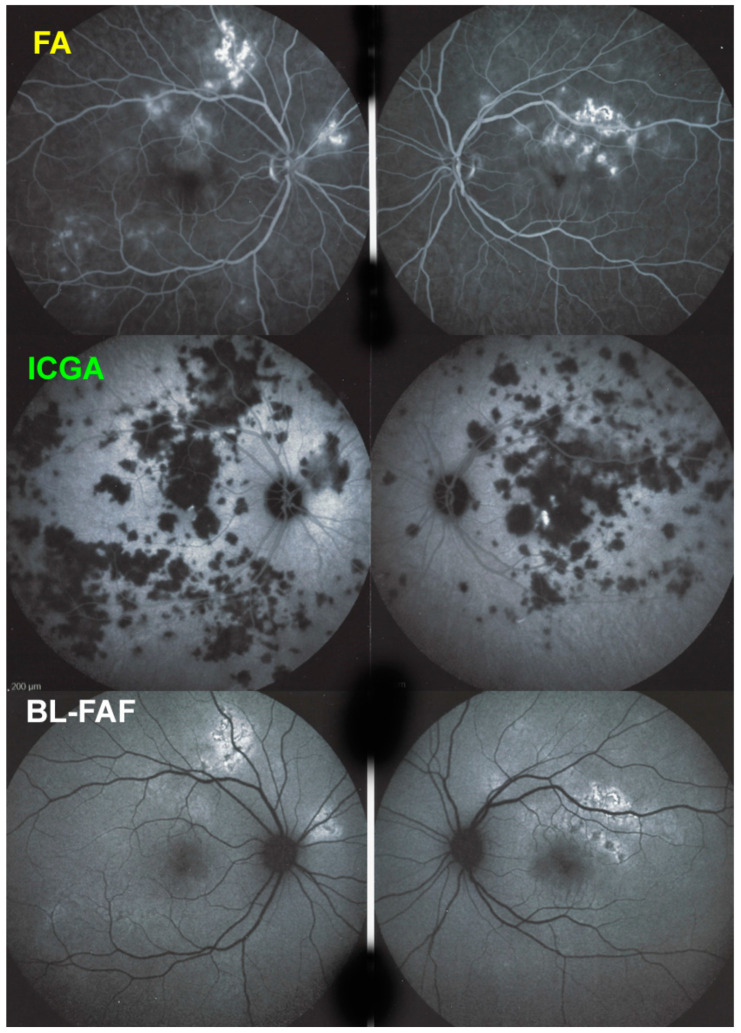
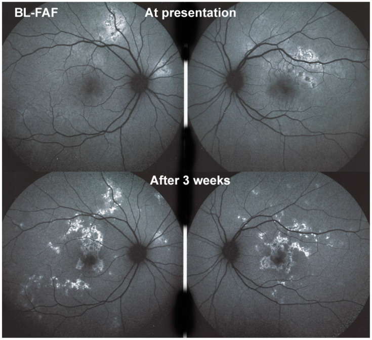
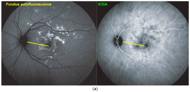
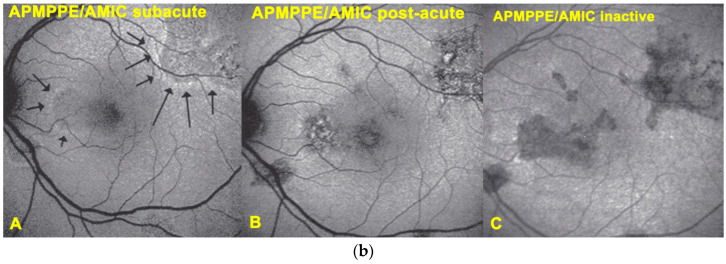
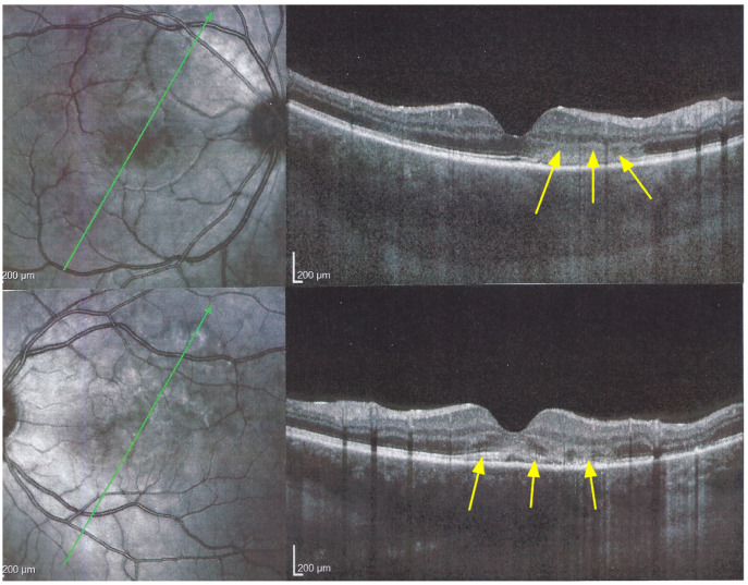
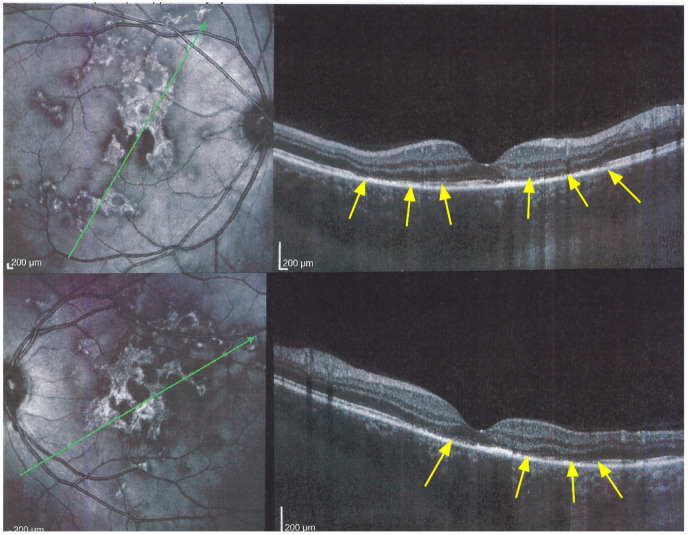
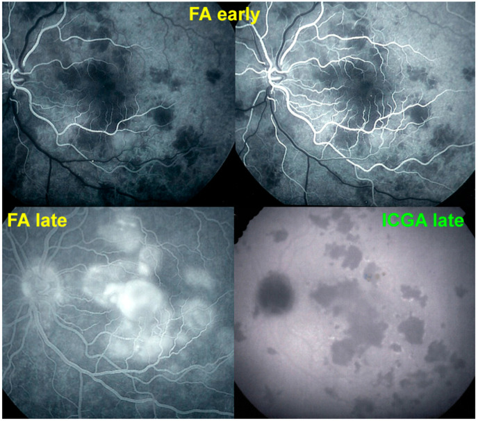
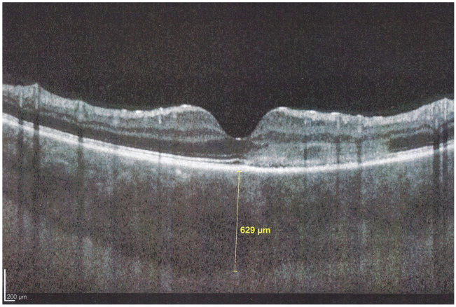
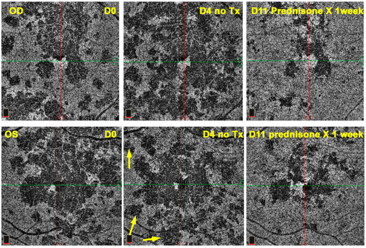
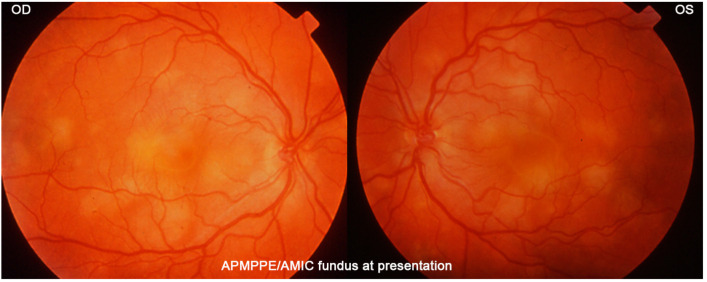
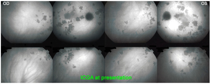
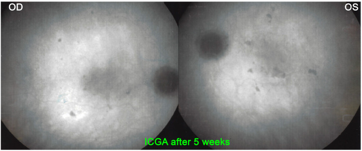
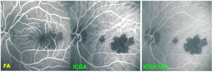
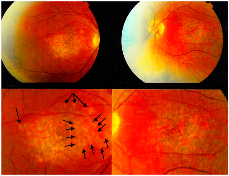
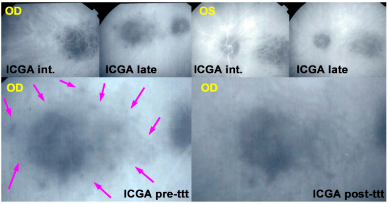
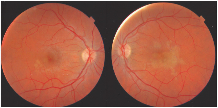
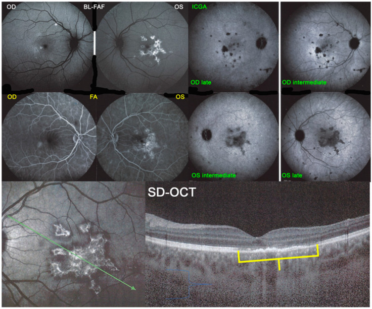
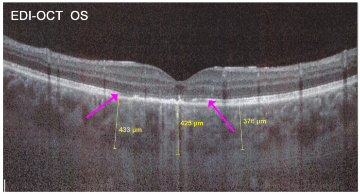
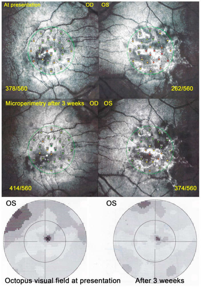
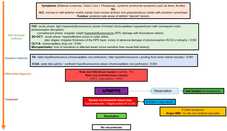
Background and objectives: Acute posterior multifocal pigment epitheliopathy/acute multifocal ischaemic choriocapillaritis (APMPPE/AMIC) is part of the group of choriocapillaritis entities. The aim of this article was to report a series of patients with emphasis on the clinical presentation and treatment paradigms. Materials and Methods: Retrospective case series study performed in the Centre for Ophthalmic Specialised care (COS), Lausanne, Switzerland, on patients diagnosed from 2000 to 2021 with APMPPE/AMIC. Procedures performed at presentation and upon follow-up (when available) included best corrected visual acuity (BCVA), routine ocular examination, laser flare photometry (LFP) microperimetry (when available) and visual field testing. Imaging investigations included spectral domain optical coherence tomography (SD-OCT)/enhanced depth imaging OCT (EDI-OCT), OCT angiography (OCT-A) as well as fluorescein and indocyanine green angiography (FA, ICGA). The presence or not of prodromal systemic viral-like symptoms was noted. The localisation of lesions whether foveal or extrafoveal, divided the patients into 2 groups (foveal, peri-or parafoveal). Exclusion criteria were patients diagnosed with APMPPE/AMIC and a positive QuantiFERON test and/or VDRL-TPHA tests. Results: Nineteen (35 eyes) of 1664 new patients (1.14%) were diagnosed with APMPPE/AMIC and included in our study. 13 (68%) were male and 6 (32%) were female. The mean age was 33.1 ± 9.2 years. 16 (84%) patients mentioned a viral prodromal episode or other systemic symptoms, and 3 (16%) did not mention any episode before the onset of ocular symptoms. 15 (39%) out of 38 eyes had foveal localisation of the lesions, 20 (52.6%) had peri- or para-foveal localisations and 3 eyes were normal [3 unilateral cases (15%)]. Mean BCVA at presentation was 0.83 ± 0.24 for the whole group. It was 0.58 ± 0.28 for the group with foveal lesions, increasing to 0.97 ± 0.13 at last follow-up (p = 0.0028). For the group with extrafoveal lesions mean BCVA at presentation was 0.94 ± 0.18, improving to 1.18± 0.10 at last follow-up (p = 0.0039). 13 (68%) patients received prednisone treatment, of whom 2 (10%) received additionally at least one immunosuppressive agent, 4 (20%) patients received no treatment and in 2 patients the information was unavailable. All patients in the foveal lesion group received corticosteroid treatment except one who evolved to bilateral macular atrophy. Conclusions: APMPPE/AMIC is a primary choriocapillaritis. Although it is thought that the disease is self-limited, treatment is necessary in most cases, especially when lesions are located in the fovea.

Keywords: APMPPE/AMIC; blue light fundus autofluorescence (BL-FAF); enhanced depth imaging OCT (EDI-OCT); indocyanine green angiography (ICGA); primary inflammatory choriocapillaropathies (PICCPs); spectral domain optical coherence tomography (SD-OCT).


Conflict of interest statement

The authors declare no conflict of interest.

Figures

Figure 1
Figure 1
Clinicopathology of PICCPs according to vessel location of non-perfusion. Schematic drawing of the different levels of choriocapillaris vessel the occlusions of which determine the type of choriocapillaritis. (Reproduced from Papasavvas I, Herbort CP Jr. Diagnosis and Treatment of Primary Inflammatory Choriocapillaropathies (PICCPs): A Comprehensive Overview. Medicina (Kaunas). 2022 Jan 21;58(2):165. doi: 10.3390/medicina58020165. PMID: 35208488; PMCID: PMC8879059).
Figure 2
Figure 2
Clinicopathology of PICCPs according to the suspected type and extension of vessel non-perfusion. Non-perfusion of low-flow end capillary vessels causes the benign and reversible disease MEWDS. If larger choriocapillaris vessels or precapillary vessels are occluded the more severe diseases develop including APMPPE, MFC and SC [7]).
Figure 3
Figure 3
APMPPE/AMIC: Bilateral fundus pictures of a patient showing the characteristic placoid yellow lesions in the posterior pole and mid-periphery following a severe febrile episode with gastroenteritis.
Figure 4
Figure 4
(a) APMPPE/AMIC: ICGA in a patient showing bilaterally scattered geographical partly confluent placoid hypofluorescent areas caused by choriocapillaris non-perfusion and corresponding visual field scotomas (inset). (b) APMPPE/AMIC: ICGA ODS in a case with limited areas of hypofluorescence (top two frames) that resolved without treatment after 7 weeks (bottom frames).
Figure 5
Figure 5
APMPPE/AMIC; FA & ICGA & BL-FAF at presentation. Case of acute APMPPE/AMIC analysed by ICGA & FA & BL-FAF. This multimodal imaging shows that the initial event is choriocapillaris non-perfusion clearly shown by extensive areas of ICGA hypofluorescence (middle two frames) while retinal (FA) involvement is still limited. BL-FAF hyperautofluorescence is still limited as choriocapillaris non-perfusion induced ischaemia did not alter the outer retina yet, except in a few areas along the superior temporal arcades.
Figure 6
Figure 6
APMPPE/AMIC; BL-FAF at presentation and in the subacute phase. At presentation (top two frames) there is limited hyperautofluorescence as there is mainly thickening of the outer retina due to choriocapillaris non-perfusion (see Figure 8) and limited areas (yet) of loss of photoreceptor outer segments (see Figure 7). During the subacute phase (bottom two frames) there are extended irregular areas of hyperfluorescence corresponding either to loss of photoreceptor outer segments allowing to see the hyperautofluorescent RPE lipofuscin or RPE cell damage with an accumulation of cellular fluorophore debris or both.
Figure 7
Figure 7
(a) APMPPE/AMIC; BL-FAF patterns in subacute stage (same patient as Figure 5 and Figure 6). Severe case after 3 weeks of evolution. BL-FAF shows central hypoautofluorescence corresponding to atrophy with loss of RPE, co-localising with dark ICGA hypofluorescence (yellow callipers), surrounded by hyperautofluorescence corresponding to non-perfusion with intact RPE and loss of photoreceptor segments and/or damaged RPE cells with accumulation of fluorophore debris. (Same patient as Figure 5 and Figure 6). (b) APMPPE/AMIC; BL-FAF evolution during stages of the disease. In the subacute stage (A) hyperautofluorescence is mainly present at the border of non-perfused areas (black arrows) but less so in the middle of diseased areas, indicating mostly non-functioning RPE cells while at the rim RPE cells are still metabolically active and lipofuscins can be seen through areas that have lost photoreceptor outer segments. At a later stage (B), hyperautofluorescent rims around the affected have disappeared because of healed RPE cells and reconstitution of the photoreceptor outer segment line. In the inactive healed phase (C) the chorioretinal scars where RPE cells have disappeared are hypoautofluorescent (Reprinted from Gupta A, Gupta V, Herbort CP, Khairallah M, editors. Uveitis, text and imaging. New Delhi: Jaypee Brothers Medical Publishers; 2009:328).
Figure 7
Figure 7
(a) APMPPE/AMIC; BL-FAF patterns in subacute stage (same patient as Figure 5 and Figure 6). Severe case after 3 weeks of evolution. BL-FAF shows central hypoautofluorescence corresponding to atrophy with loss of RPE, co-localising with dark ICGA hypofluorescence (yellow callipers), surrounded by hyperautofluorescence corresponding to non-perfusion with intact RPE and loss of photoreceptor segments and/or damaged RPE cells with accumulation of fluorophore debris. (Same patient as Figure 5 and Figure 6). (b) APMPPE/AMIC; BL-FAF evolution during stages of the disease. In the subacute stage (A) hyperautofluorescence is mainly present at the border of non-perfused areas (black arrows) but less so in the middle of diseased areas, indicating mostly non-functioning RPE cells while at the rim RPE cells are still metabolically active and lipofuscins can be seen through areas that have lost photoreceptor outer segments. At a later stage (B), hyperautofluorescent rims around the affected have disappeared because of healed RPE cells and reconstitution of the photoreceptor outer segment line. In the inactive healed phase (C) the chorioretinal scars where RPE cells have disappeared are hypoautofluorescent (Reprinted from Gupta A, Gupta V, Herbort CP, Khairallah M, editors. Uveitis, text and imaging. New Delhi: Jaypee Brothers Medical Publishers; 2009:328).
Figure 8
Figure 8
APMPPE/AMIC; SD-OCT in initial-acute phase OD (top) and OS (bottom) (same patient as Figure 5, Figure 6 and Figure 7a). In this initial phase, SD-OCT shows areas of hyperreflective changes visible in the outer plexiform layer and adjacent outer nuclear layer (arrows) but no loss of photoreceptor outer segments (see Figure 5).
Figure 9
Figure 9
APMPPE/AMIC; SD-OCT in subacute phase OD (top) and OS (bottom) (same patient as Figure 5, Figure 6, Figure 7a and Figure 8). In the subacute phase, SD-OCT shows extended areas of loss of photoreceptor outer segments (arrows) and thickened RPE, hyperautofluorescent on BL-FAF (see Figure 5).
Figure 10
Figure 10
Late FA pooling in a severe case of APMPPE/AMIC. In severe cases of APMPPE/AMIC, FA shows choriocapillaris non-perfusion in early angiographic frames (top two pictures—FA early) followed by areas of abundant retinal pooling in the late angiographic phase (bottom left picture—FA late). Traditionally this phenomenon is explained by the very hypothetical alleged change of polarity of the RPE and fluid movement from the choroid to the retina. However, the choriocapillaris areas under the retinal pooling are non-perfused (bottom right picture—ICGA late). Therefore, a more probable origin of the fluid is an exudation from retinal vessels in response to severe outer retinal ischemia.
Figure 11
Figure 11
APMPPE/AMIC; EDI-OCT in the acute phase (same patient as Figure 5, Figure 6, Figure 7a, Figure 8 and Figure 9). In the acute phase, EDI-OCT shows an important thickening of the whole choroid with the calliper indicating a thickness of 629 µm.
Figure 12
Figure 12
APMPPE/AMIC; OCT-angiography (OCT-A). OCT-A shows the choriocapillary drop-out at presentation (top left frame) and its progressive decrease up to 3 months after the onset of the disease (bottom right frame).
Figure 13
Figure 13
APMPPE/AMIC; Microperimetry (MP). MP of a case of APMPPE/AMIC at presentation (top two pictures) showing a decrease of retinal sensitivity to 268/550 (OD) and 95/560, recovering to 448/560 (OD) and 438/560 (OS) after 6 months (bottom two pictures).
Figure 14
Figure 14
APMPPE/AMIC SD-OCT; Subretinal fluid producing serous retinal detachment (SRDs) and intra-photoreceptor layer separation, in a hyperacute case.
Figure 15
Figure 15
APMPPE/AMIC SD-OCT; Chorioretinal atrophy in the late stage.
Figure 16
Figure 16
APMPPE/AMIC OCT-A; Case of hyperacute APMPPE/AMIC with numerous hypo-perfused areas at presentation (D0). Prednisone treatment was withheld until negative syphilis serology was available with the progression of non-perfused areas (D4 yellow arrows). One week after the introduction of systemic prednisone therapy (D11) lesions had regressed substantially.
Figure 17
Figure 17
APMPPE/AMIC: Typical case of a 27-year-old patient showing the characteristic yellow-white placoid fundus lesions.
Figure 18
Figure 18
APMPPE/AMIC, ICGA at presentation: Characteristic bilateral hypofluorescent areas representing choriocapillaris non-perfusion.
Figure 19
Figure 19
APMPPE/AMIC, ICGA after 5 weeks: Resolution of most of the hypofluorescent areas after systemic prednisone treatment.
Figure 20
Figure 20
APMPPE/AMIC at first presentation elsewhere. FA early frame (left) shows two areas of non-perfusion, which are better delineated by the ICGA intermediate phase (middle frame) and late phase (right frame).
Figure 21
Figure 21
APMPPE/AMIC case not having received prednisone therapy with an evolution towards macular atrophy ODS: This patient presented with macular atrophy OS following involvement first of the left eye and placoid lesions in the right fundus still partially active (black arrows).
Figure 22
Figure 22
APMPPE/AMIC; ICGA in a case that did not receive prednisone therapy with an evolution towards macular atrophy ODS: top frames show ICGA findings in intermediate and late phases in both eyes at presentation 3 months after the onset of disease. The bottom left frame shows active perimacular lesions less hypofluorescent than the centre (crimson arrows) that responded to prednisone therapy with regression of perimacular lesions after treatment but the persistence of centromacular atrophy (bottom right frame).
Figure 23
Figure 23
APMPPE/AMIC case at a subacute stage having received prednisone therapy for 10 days. Faint placoid lesions still visible OS and faint macular discolouration OD.
Figure 24
Figure 24
APMPPE/AMIC; BL-FAF. ICGA, FA & SD-OCT findings in a case in subacute stage having received prednisone therapy for 10 days: BL-FAF (top left two frames) shows areas of hyperautofluorescence (OS > OD) corresponding to the areas of hypofluorescence (choriocapillaris non-perfusion) on ICGA (top right quartet of frames). FA (middle left two frames) shows areas of staining (OS) most probably produced by exudation of retinal vessels in response to outer retinal ischaemia. SD-OCT (bottom two pictures) shows loss and disorganisation of photoreceptor outer segments, disrupted RPE layer with overlying hyperreflective focal deposits in correspondence to hyperautofluorescence.
Figure 25
Figure 25
APMPPE/AMIC; EDI-OCT (same case as Figure 21 and Figure 22). Besides the loss of photoreceptor outer segments (marked by crimson arrows), the whole choroid is thickened, with callipers showing values between 376 µm and 433 µm.
Figure 26
Figure 26
APMPPE/AMIC; Microperimetry and visual field evolution (same patient as Figure 21, Figure 22 and Figure 23): Microperimetry is closely and numerically monitoring the evolution of retinal sensitivity from 373/560 to 414/560 OD and from 262/560 to 374/560 OS and is much more precise than visual field testing (bottom 2 images).
Scheme 1
Scheme 1
APMPPE/AMIC: decision tree in clinical practice.

Similar articles

Cited by

References

    1. Gass J.D.M. Acute Posterior Multifocal Placoid Pigment Epitheliopathy. Arch. Ophthalmol. 1968;80:171–185. doi: 10.1001/archopht.1968.00980050179005. - DOI - PubMed
    1. Deutman A.F., Oosterhuis J.A., Boen-Tan T.N., Aan de Kerk A.L. Acute posterior multifocal placoid pigment epitheliopathy. Pigment epitheliopathy or choriocapillaritis. Br. J. Ophthalmol. 1972;56:863–874. doi: 10.1136/bjo.56.12.863. - DOI - PMC - PubMed
    1. Deutman A.F., Lion F. Choriocapillaris non-perfusion in acute multifocal placoid pigment epitheliopathy. Am. J. Ophthalmol. 1977;84:652–657. doi: 10.1016/0002-9394(77)90380-4. - DOI - PubMed
    1. Howe L.J., Woon H., Graham E.M., Fitzke F., Bhandari A., Marshall J. Choroidal hypoperfusion in acute posterior multifocal placoid pigment epitheliopathy. An indocyanine green angiography study. Ophthalmology. 1995;102:790–798. doi: 10.1016/S0161-6420(95)30955-4. - DOI - PubMed
    1. Herbort C.P., Jr., Mantovani A., Tugal-Tutkun I., Papasavvas I. Classification of Non-Infectious and/or Immune Mediated Choroiditis: A Brief Overview of the Essentials. Diagnostics. 2021;11:939. doi: 10.3390/diagnostics11060939. - DOI - PMC - PubMed

LinkOut - more resources