

Human Cytomegalovirus and Human Herpesvirus 6 Coinfection of Dermal Fibroblasts Enhances the Pro-Inflammatory Pathway Predisposing to Fibrosis: The Possible Impact on Systemic Sclerosis
- PMID: 36014018
- PMCID: PMC9415275
- DOI: 10.3390/microorganisms10081600
Human Cytomegalovirus and Human Herpesvirus 6 Coinfection of Dermal Fibroblasts Enhances the Pro-Inflammatory Pathway Predisposing to Fibrosis: The Possible Impact on Systemic Sclerosis
Abstract
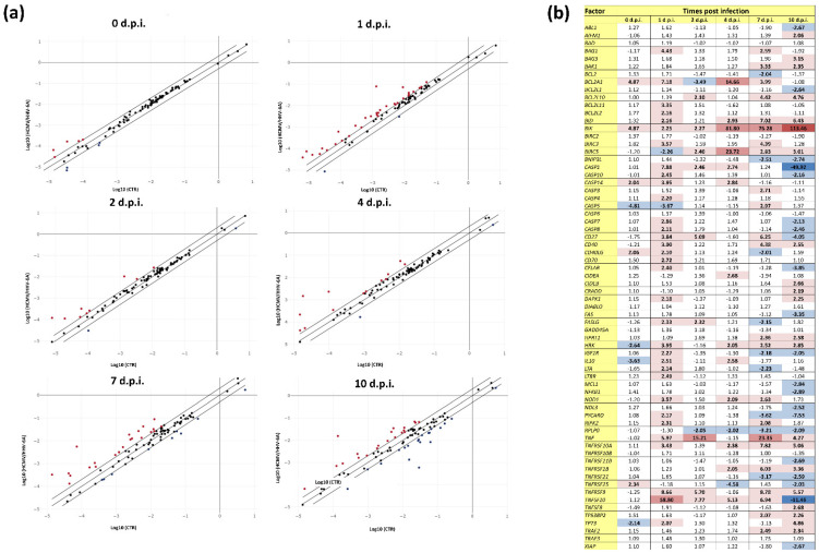
Systemic sclerosis (SSc) is a severe autoimmune disease likely triggered by genetic and environmental factors, including viral infections. Human cytomegalovirus (HCMV) and human herpesvirus 6A species (HHV-6A) have been associated with SSc, based on in vivo and in vitro evidence, but the data are still inconclusive. Furthermore, despite both viruses being highly prevalent in humans and able to exacerbate each other's effects, no data are available on their joint effects. Hence, we aimed to study their simultaneous impact on the expression of cell factors correlated with fibrosis and apoptosis in in vitro coinfected fibroblasts, representing the main target cell type in SSc. The results, obtained by a microarray detecting 84 fibrosis/apoptosis-associated factors, indicated that coinfected cells underwent higher and more sustained expression of fibrosis-associated parameters compared with single-infected cells. Thus, the data, for the first time, suggest that HCMV and HHV-6A may cooperate in inducing alterations potentially leading to cell fibrosis, thus further supporting their joint role in SSc. However, further work is required to definitively answer whether β-herpesviruses are causally linked to the disease and to enable the possible use of targeted antiviral treatments to improve clinical outcomes.
Keywords: HCMV; HHV-6; systemic sclerosis; tissue fibrosis factors.
Conflict of interest statement
The authors declare no conflict of interest.
Figures


Similar articles
-
Virus-Induced MicroRNA Modulation and Systemic Sclerosis Disease.Biomedicines. 2024 Jun 19;12(6):1360. doi: 10.3390/biomedicines12061360. Biomedicines. 2024. PMID: 38927567 Free PMC article. Review.
-
Coinfection of Dermal Fibroblasts by Human Cytomegalovirus and Human Herpesvirus 6 Can Boost the Expression of Fibrosis-Associated MicroRNAs.Microorganisms. 2023 Feb 6;11(2):412. doi: 10.3390/microorganisms11020412. Microorganisms. 2023. PMID: 36838377 Free PMC article.
-
Impact of Human Cytomegalovirus and Human Herpesvirus 6 Infection on the Expression of Factors Associated with Cell Fibrosis and Apoptosis: Clues for Implication in Systemic Sclerosis Development.Int J Mol Sci. 2020 Sep 3;21(17):6397. doi: 10.3390/ijms21176397. Int J Mol Sci. 2020. PMID: 32899126 Free PMC article.
-
Modulation of microRNome by Human Cytomegalovirus and Human Herpesvirus 6 Infection in Human Dermal Fibroblasts: Possible Significance in the Induction of Fibrosis in Systemic Sclerosis.Cells. 2021 Apr 29;10(5):1060. doi: 10.3390/cells10051060. Cells. 2021. PMID: 33946985 Free PMC article.
-
Insights into the knowledge of complex diseases: Environmental infectious/toxic agents as potential etiopathogenetic factors of systemic sclerosis.J Autoimmun. 2021 Nov;124:102727. doi: 10.1016/j.jaut.2021.102727. Epub 2021 Oct 1. J Autoimmun. 2021. PMID: 34601207 Review.
Cited by
-
Virus-Induced MicroRNA Modulation and Systemic Sclerosis Disease.Biomedicines. 2024 Jun 19;12(6):1360. doi: 10.3390/biomedicines12061360. Biomedicines. 2024. PMID: 38927567 Free PMC article. Review.
-
Circulating miRNAs Expression in Myalgic Encephalomyelitis/Chronic Fatigue Syndrome.Int J Mol Sci. 2023 Jun 24;24(13):10582. doi: 10.3390/ijms241310582. Int J Mol Sci. 2023. PMID: 37445763 Free PMC article.
-
Coinfection of Dermal Fibroblasts by Human Cytomegalovirus and Human Herpesvirus 6 Can Boost the Expression of Fibrosis-Associated MicroRNAs.Microorganisms. 2023 Feb 6;11(2):412. doi: 10.3390/microorganisms11020412. Microorganisms. 2023. PMID: 36838377 Free PMC article.
References
-
- Ferri C., Arcangeletti M.C., Caselli E., Zakrzewska K., Maccari C., Calderaro A., D’Accolti M., Soffritti I., Arvia R., Sighinolfi G., et al. Insights into the knowledge of complex diseases: Environmental infectious/toxic agents as potential etiopathogenetic factors of systemic sclerosis. J. Autoimmun. 2021;124:102727. doi: 10.1016/j.jaut.2021.102727. - DOI - PubMed
Grants and funding
LinkOut - more resources
Full Text Sources
