Review
. 2022 Aug 17;10(8):1659.
doi: 10.3390/microorganisms10081659.

Anti- Toxoplasma gondii IgM Long Persistence: What Are the Underlying Mechanisms?

Affiliations
Review

Anti- Toxoplasma gondii IgM Long Persistence: What Are the Underlying Mechanisms?

José Antonio Vargas-Villavicencio et al. Microorganisms. .

Abstract

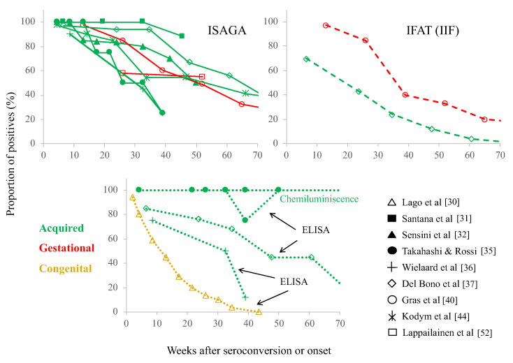
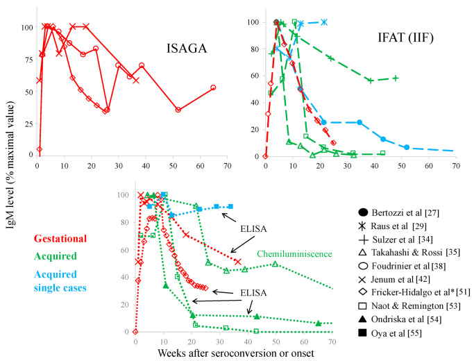
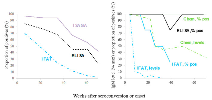
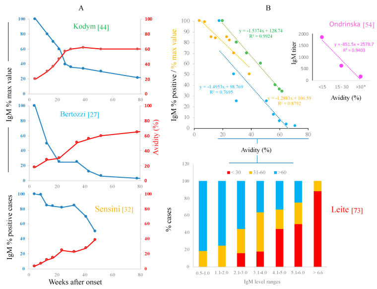
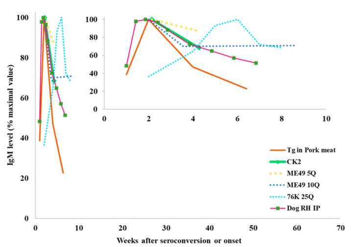
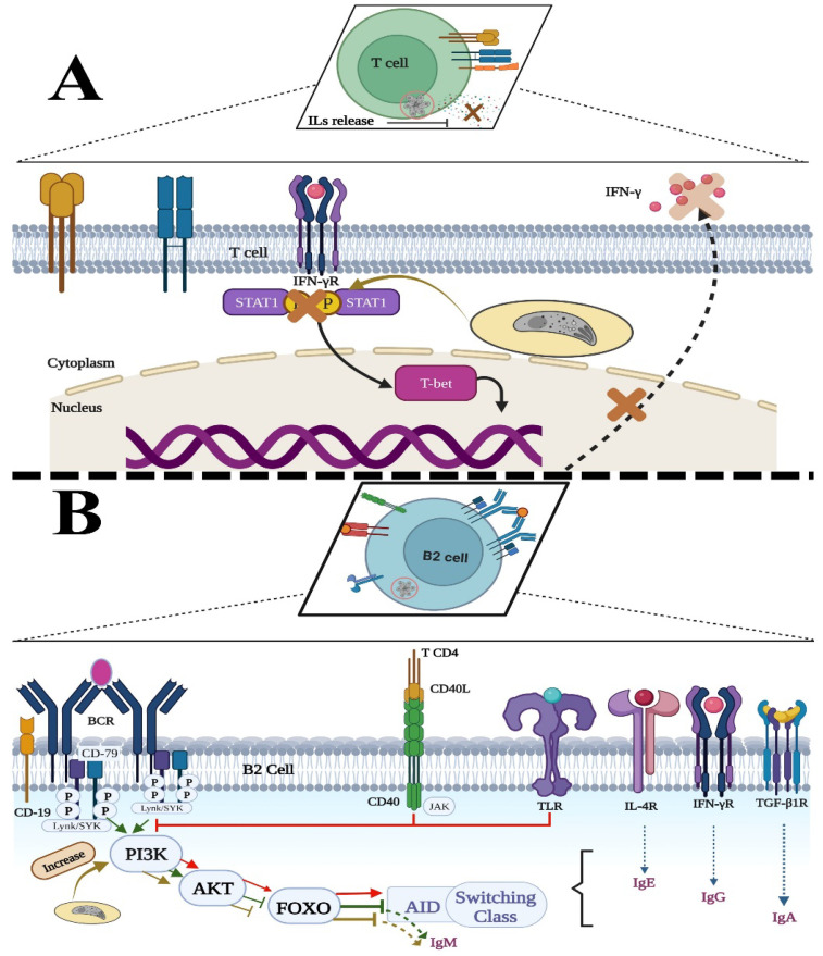
Diagnosis of Toxoplasma gondii acute infection was first attempted by detection of specific IgM antibodies, as for other infectious diseases. However, it was noted that this immunoglobulin declines slowly and may last for months or even years. Apart from the diagnostic problem imposed on clinical management, this phenomenon called our attention due to the underlying phenomena that may be causing it. We performed a systematic comparison of reports studying IgM antibody kinetics, and the data from the papers were used to construct comparative plots and other graph types. It became clear that this phenomenon is quite generalized, and it may also occur in animals. Moreover, this is not a technical issue, although some tests make more evident the prolonged IgM decay than others. We further investigated biological reasons for its occurrence, i.e., infection dynamics (micro-reactivation-encystment, reinfection and reactivation), parasite strain relevance, as well as host innate, natural B cell responses and Ig class-switch problems inflicted by the parasite. The outcomes of these inquiries are presented and discussed herein.

Keywords: IgM; Toxoplasma gondii; serological diagnosis.


Conflict of interest statement

The authors declare no conflict of interest.

Figures

Figure 1
Figure 1
Immune response against Toxoplasma gondii. Both gestational and acquired toxoplasmosis occur by the ingestion of tissue cysts present in undercooked meat or oocysts that come from felines that release them with their feces; sometimes, water and legumes/pastures are contaminated. Thus, the parasite reaches the digestive tract. The sporozoite or bradyzoite released from the oocyst or tissue cyst, respectively, invades the enterocyte or crosses the epithelium to reach the submucosa, where there are gut-associated lymphoid tissues (GALT, such as Peyer’s patches) where a response begins: (a) Local macrophages (Mo) phagocytize the parasite and initiate an inflammatory response, together with local granulocytes (neutrophils, Ne); (b) B-1 cells can produce IgM and IgG antibodies (and IgA) after antigen or Pathogen-associated Molecular Patterns (PAMPs) recognition through the B cell Receptor (BCR), the Pattern Recognition Receptors (PRRs)- mainly Toll-Like Receptors (TLRs)- or both; B-1 and some marginal zone B cells (MZB) are able to spontaneously produce “natural” antibodies to microbiota, pathogens or autoantigens, that may cross-react with T. gondii; (c) Dendritic cells (DCs) take up antigens and travel to the nearest lymph node (LN) guided by chemokines recognized by specific receptors (such as CXCR5); at the LN, they stimulate both T helper lymphocytes (Th-CD4+), which convert into Th1 and produce Interferon-gamma (IFN-γ) and other cytokines, and cytotoxic T cells (CTL, CD8+), which become important for infected cell destruction; (d) The antigen also reaches the LN where the B-2 (follicular) B cells are primed and become available for CD4+ T help, undergoing antibody class switch. After selection of highly specific clones by DCs, they become plasma cells (PCs), which migrate to the bone marrow (BM) and secrete IgG, IgA or IgE antibodies. B-2, CD4+ and CD8+ memory cells are also born in the LN and migrate to different tissues, where they react more rapidly to a second stimulus. Protective mechanisms include complement fixation, opsonized phagocytosis, infected cell cytotoxicity by IFN-γ activated NKs and CTLs. This response is modulated by regulatory T lymphocytes and other cell types, including B-1 (B10) and M2 macrophages, directly or through tumor growth factor beta (TGF-β) or Interleukin (IL)-10. This summary is oversimplified to direct attention to antibody production. Please refer to relevant reviews of this topic for further details and other cell types and molecules involved [6,7,8,9,10,11,12]. Graphical contents were created with BioRender.com (license MX249HDDR2, accessed on 9 August 2022).
Figure 2
Figure 2
Proportion of cases positive for T. gondii reacting IgM antibodies over time in different groups after initial clinical or serological signs of infection [30,31,32,35,36,37,40,44,52]. Each graph corresponds to the test employed, as labelled. ISAGA: Immunosorbent Agglutination Assay; IFAT: Indirect Immunofluorescence Antibody Test (some articles refer to this technique as IIF or IFI); ELISA (EIA): Enzyme-linked Immunosorbent Assay; Chemiluminescence: microbead-based automated test. The abscissa axis cuts off at 70 weeks, but several studies followed the patients for longer periods.
Figure 3
Figure 3
Kinetics of IgM antibody levels reactive to T. gondii in different groups after initial clinical or serological signs of infection [27,29,34,35,38,42,51,53,54,55]. The IgM levels were normalized to percentage of the maximum value in order to compare among studies. Dispersion data are not displayed (even if reported). The plots are separated according to the test performed. Acronyms of tests are as in Figure 2. The x-axis also cuts off at 70 weeks, but two studies followed patients for longer periods. * Fricker-Hidalgo et al. compared techniques, but we could only gather data from ISAGA, IFAT and ELISA.
Figure 4
Figure 4
Kinetics of IgM decline assessed by different techniques and data presentation form (proportion of positives versus antibody levels) in two studies that compared these parameters with the same samples [35,37]. In both works, the patients had clinically active acquired infection.
Figure 5
Figure 5
Relationship between IgM and avidity as seen in kinetics (A) or direct relation/proportion of cases (B). Data from tables or plots of the original works [27,32,44,54,73] were taken and used to build the graphs. Names in different colors are the first authors of these reports. * Considered chronic in that study.
Figure 6
Figure 6
Kinetics of anti-T. gondii-reacting IgM antibody levels in animal groups experimentally infected. The large graphic shows the kinetics until 70 weeks in order to compare with human cases. The inset shows a shorter period amplified [75,76,77,78,79] and adjusted to percent of maximal value, with the purpose of normalizing and comparing among studies. CK2 and RH: virulent strains; ME49 and 76K: non-virulent strains; Tg: T. gondii; #Q: number of cysts inoculated; IP: intraperitoneal.
Figure 7
Figure 7
Possible mechanisms by which IgM can be prolonged: (A) Toxoplasma can infect CD4+ T lymphocytes. Within the cell, it could inhibit the phosphorylation of several receptors, resulting in minimal production of cytokines necessary for secondary stimulus in class switching. (B) Toxoplasma may invade B cells and enhances phosphatidyl inositol 3 kinase (PI3K) to levels that stimulate protein kinase B (AKT), inhibiting fork-head box O transcription factor (FOXO) and, consequently, the activation-induced cytidine deaminase (AID), a key enzyme for class-switch recombination (CSR). STAT-1: transducer and activator of transcription protein-1; T-bet: T cell transcription factor; BCR: B cell receptor; TLR: Toll-Like Receptor; IL-4R, IFN-γR and TGF-βR: Receptors for the cytokines IL-4, IFN-γ and TGF-β. Graphical contents were created with BioRender.com (licenses ZC249HCMVZ and PE249HCGKN, accessed on 9 August 2022).

Similar articles

Cited by

References

    1. Montoya J.G., Liesenfeld O. Toxoplasmosis. Lancet. 2004;363:1965–1976. doi: 10.1016/S0140-6736(04)16412-X. - DOI - PubMed
    1. Dubey J.P. History of the Discovery of the life cycle of Toxoplasma gondii. Int. J. Parasitol. 2009;39:877–882. doi: 10.1016/j.ijpara.2009.01.005. - DOI - PubMed
    1. Hall S.M., Surveillance D. Congenital toxoplasmosis. BMJ. 1992;305:291–297. doi: 10.1136/bmj.305.6848.291. - DOI - PMC - PubMed
    1. Dunn D., Wallon M., Peyron F., Petersen E., Peckham C., Gilbert R. Mother-to-child transmission of toxoplasmosis: Risk estimates for clinical counselling. Lancet. 1999;353:1829–1833. doi: 10.1016/S0140-6736(98)08220-8. - DOI - PubMed
    1. Suzuki Y., Sa Q., Ochiai E., Mullins J., Yolken R., Halonen S.K. Toxoplasma gondii Model Apicomplexan Perspectives and Methods. 2nd ed. Elsevier Ltd.; Amsterdam, The Netherlands: 2014. Toxoplasma gondii; pp. 755–796. - DOI

LinkOut - more resources