Review
. 2022 Aug 1;13(8):1238.
doi: 10.3390/mi13081238.

Microfluidics-Based POCT for SARS-CoV-2 Diagnostics

Affiliations
Review

Microfluidics-Based POCT for SARS-CoV-2 Diagnostics

Binfeng Yin et al. Micromachines (Basel). .

Abstract

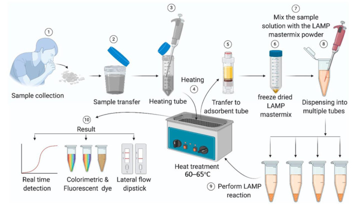
A microfluidic chip is a tiny reactor that can confine and flow a specific amount of fluid into channels of tens to thousands of microns as needed and can precisely control fluid flow, pressure, temperature, etc. Point-of-care testing (POCT) requires small equipment, has short testing cycles, and controls the process, allowing single or multiple laboratory facilities to simultaneously analyze biological samples and diagnose infectious diseases. In general, rapid detection and stage assessment of viral epidemics are essential to overcome pandemic situations and diagnose promptly. Therefore, combining microfluidic devices with POCT improves detection efficiency and convenience for viral disease SARS-CoV-2. At the same time, the POCT of microfluidic chips increases user accessibility, improves accuracy and sensitivity, shortens detection time, etc., which are beneficial in detecting SARS-CoV-2. This review shares recent advances in POCT-based testing for COVID-19 and how it is better suited to help diagnose in response to the ongoing pandemic.

Keywords: SARS-CoV-2; microfluidic; point of care testing.


Conflict of interest statement

The authors declare no conflict of interest.

Figures

Figure 1
Figure 1
Schematic view of POCT methods for microfluidic use to detect SARS-CoV-2.
Figure 2
Figure 2
(a) Photo of a portable centrifugal microfluidic device. (b) Schematic diagram of each component of the microfluidic device. (c) Schematic diagram of the structure and functional areas of the centrifugal microfluidic chip. (Reprinted/adapted with permission from Ref. [34]. Copyright 2020 Royal Society of Chemistry).
Figure 3
Figure 3
Schematic diagram of the LAMP detection process [45].
Figure 4
Figure 4
(a) A microfluidic chip structure of sequential fluid dispensing. (Reprinted/adapted with permission from Ref. [46]. Copyright 2021 Royal Society of Chemistry). (b) A microfluidic chip integrated LAMP and particle diffusometry. (Reprinted/adapted with permission from Ref. [49]. Copyright 2022 Elsevier).
Figure 5
Figure 5
(a) Overview of the universally stable and precise CRISPR-LAMP detection platform. (Reprinted/adapted with permission from Ref. [59]. Copyright 2020 American Chemical Society). (b) A lateral flow microfluidic device that does not rely on external devices and has a hand-warmer pouch as its power source. (Reprinted/adapted with permission from Ref. [62]. Copyright 2022 Elsevier).
Figure 6
Figure 6
(a) Screening and optimization of the ssDNA-FQ reporters for RAVI-CRISPR assays. (Reprinted/adapted with permission from Ref. [91]. Copyright 2022 American Chemical Society). (b) Schematic for the smartphone antibody detection workflow. (Reprinted/adapted with permission from Ref. [92]. Copyright 2021 American Chemical Society).
Figure 7
Figure 7
Structure and flow diagram of colorimetric lateral flow immunoassay. (Reprinted/adapted with permission from Ref. [102]).
Figure 8
Figure 8
(a) Graphical illustration of the step-by-step immuno-biosensor preparation. (Reprinted/adapted with permission from Ref. [113]. Copyright 2020 American Chemical Society). (b) Schematic diagram of electrochemical biosensor detection of a saliva sample. (Reprinted/adapted with permission from Ref. [115]. Copyright 2020 Elsevier).

Similar articles

Cited by

References

    1. Kim D., Lee J.Y., Yang J.S., Kim J.W., Kim V.N., Chang H. The Architecture of SARS-CoV-2 Transcriptome. Cell. 2020;181:914–921. doi: 10.1016/j.cell.2020.04.011. - DOI - PMC - PubMed
    1. Guo Y.R., Cao Q.D., Hong Z.S., Tan Y.Y., Chen S.D., Jin H.J., Tan K.S., Wang D.Y., Yan Y. The origin, transmission and clinical therapies on coronavirus disease 2019 (COVID-19) outbreak—An update on the status. Military Med. Res. 2020;7:11. doi: 10.1186/s40779-020-00240-0. - DOI - PMC - PubMed
    1. Jin Y.H., Cai L., Cheng Z.S., Cheng H., Deng T., Fan Y.P., Fang C., Huang D., Huang L.Q., Huang Q., et al. A rapid advice guideline for the diagnosis and treatment of 2019 novel coronavirus (2019-nCoV) infected pneumonia (standard version) Mil. Med. Res. 2020;7:4. doi: 10.1186/s40779-020-0233-6. - DOI - PMC - PubMed
    1. Zheng Y.Y., Ma Y.T., Zhang J.Y., Xie X. COVID-19 and the cardiovascular system. Nat. Rev. Cardiol. 2020;17:259–260. doi: 10.1038/s41569-020-0360-5. - DOI - PMC - PubMed
    1. Augustin M.N., Mbeva J.B.K. Knowledge, attitudes, and behaviors of healthcare professionals at the start of an Ebola virus epidemic. Infect. Dis. Now. 2021;51:50–54. doi: 10.1016/j.medmal.2020.04.010. - DOI - PubMed