Review
. 2022 Aug 10;27(16):5089.
doi: 10.3390/molecules27165089.

Recent Advances in Age-Related Macular Degeneration Therapies

Affiliations
Review

Recent Advances in Age-Related Macular Degeneration Therapies

Marie Fabre et al. Molecules. .

Abstract

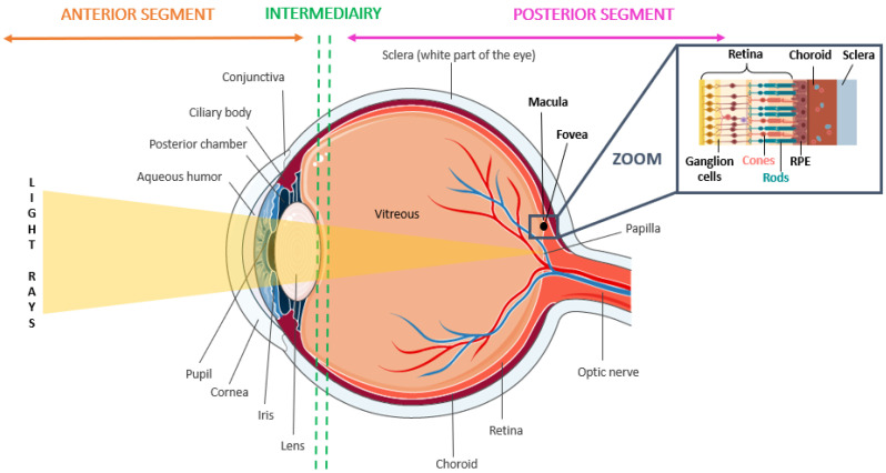
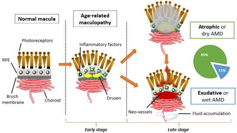
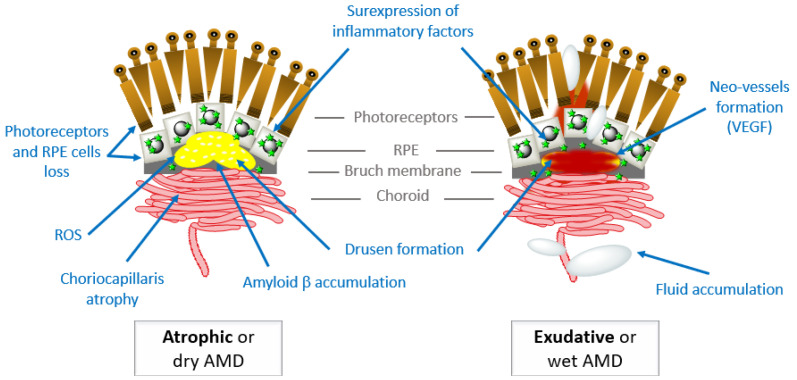
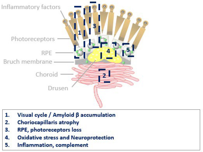
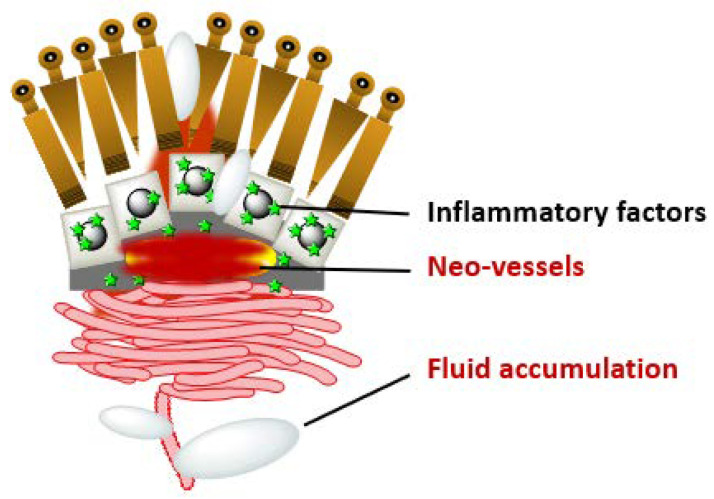
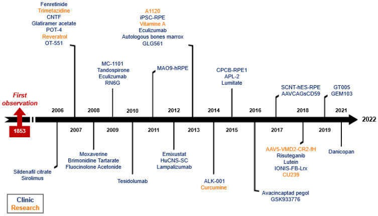
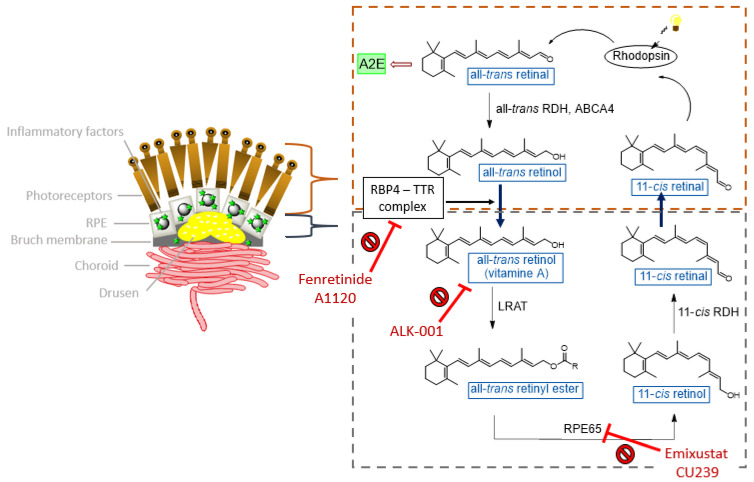
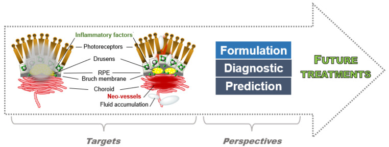
Age-related macular degeneration (AMD) was described for the first time in the 1840s and is currently the leading cause of blindness for patients over 65 years in Western Countries. This disease impacts the eye's posterior segment and damages the macula, a retina section with high levels of photoreceptor cells and responsible for the central vision. Advanced AMD stages are divided into the atrophic (dry) form and the exudative (wet) form. Atrophic AMD consists in the progressive atrophy of the retinal pigment epithelium (RPE) and the outer retinal layers, while the exudative form results in the anarchic invasion by choroidal neo-vessels of RPE and the retina. This invasion is responsible for fluid accumulation in the intra/sub-retinal spaces and for a progressive dysfunction of the photoreceptor cells. To date, the few existing anti-AMD therapies may only delay or suspend its progression, without providing cure to patients. However, in the last decade, an outstanding number of research programs targeting its different aspects have been initiated by academics and industrials. This review aims to bring together the most recent advances and insights into the mechanisms underlying AMD pathogenicity and disease evolution, and to highlight the current hypotheses towards the development of new treatments, i.e., symptomatic vs. curative. The therapeutic options and drugs proposed to tackle these mechanisms are analyzed and critically compared. A particular emphasis has been given to the therapeutic agents currently tested in clinical trials, whose results have been carefully collected and discussed whenever possible.

Keywords: age-related maculopathy; clinical trials; dry age-related macular degeneration; elderly; eye’s disease; wet age-related macular degeneration.


Conflict of interest statement

The authors declare no conflict of interest.

Figures

Figure 1
Figure 1
Anatomy of the eye divided into different segments (anterior, intermediate, and posterior) including the macula corresponding to the AMD disease area.
Figure 2
Figure 2
Incidence in Europe of the various eye diseases in people over 65 years of age [7].
Figure 3
Figure 3
Vision losses due to the advanced AMD stages.
Figure 4
Figure 4
AMD prevalence by age and gender in three studies. BEAVER DAM (American), BLUE MOUNTAINS (Australian), and EUREYE (European).
Figure 5
Figure 5
Prevalence in Europe for all ages (from <64 years to >75 years). eAMD: early AMD. nAMD: neovascular AMD. GA: geographic atrophy.
Figure 6
Figure 6
Samples of imagery commonly used to diagnose AMD. (A,B) Early AMD infra-red/OCT with serous drusen (lipidic aggregates—see text for details); significant detachment of the pigment epithelium retrofoveolar is also observed. (C) Age-related maculopathy retinography with serous drusen. (D) Atrophic AMD monitored with infra-red/OCT: macular chorioretinal atrophy ranges and external retinal atrophy with disappearance of the outer nuclear layer. (E) Exudative AMD infra-red/OCT: foveolar section showing retinal exudation with detachment of the pigment epithelium, intraretinal oedema and subretinal fluid. (F) Fluorescein angiography (left) and ICG (right) showing an exudative retro-foveolar type 1 neovessel. (G) ICG angiography showing a retro-foveolar type 1 neovessel.
Figure 7
Figure 7
The different AMD stages starting from normal macula. Age-related maculopathy (ARM), which is characterized by the appearance of drusens and inflammatory factors, then worsens in atrophic AMD and/or in exudative AMD with neovascularization and vascular leakage. RPE: retinal pigment epithelium.
Figure 8
Figure 8
Strategies and targeted elements for AMD treatments. The treatments target the loss of photoreceptors/RPE cells; ROS accumulation; choroid atrophy; inflammatory factors; β-amyloid; drusen formation; neovascularization and vascular leakage.
Figure 9
Figure 9
Current therapeutic targets for the treatment of atrophic AMD.
Figure 10
Figure 10
Current therapeutic targets for the treatment of exudative AMD.
Figure 11
Figure 11
Timeline of therapeutic agents developed against atrophic AMD.
Figure 12
Figure 12
Visual cycle in the retina with AMD disease. A2E: N-retinylidene-N-retinylethanolamine; ABCA4: ATP-Binding Cassette Subfamily A Member 4; LRAT: lecithin retinol acyltransferase; RBP4: retinol 4; RDH: retinol dehydrogenase; RPE65: retinal pigment epithelium-specific 65; TTR: transthyretin.
Figure 13
Figure 13
Lipofuscin formation.
Scheme 1
Scheme 1
Vitamin A dimerization. Dimerization occurs after cleavage of a C–H bond in position C20.
Figure 14
Figure 14
AMD progression due to ROS and inflammatory factors.
Scheme 2
Scheme 2
OT-551 transformation in the eye. The prodrug has the ability to penetrate the cornea compared to TP-H, then OT-551 is transformed in TP-H in the retina thanks to esterases.
Figure 15
Figure 15
The complement cascade. MBL: mannose binding lectin; MASP: MBL-associated serine protease; MAC: Membrane attack complex. Alternative pathway: linkage of Factor B with the hydrolyzed C3, then cleavage by the Factor D to form the C3 convertase. C3 convertase cleaves and actives the complement C3 for C3a and C3b conversion. Properdin is a positive complement to stabilize C3 and C5 convertases. Factor H intervenes over the C3/C5 dissociation and the Factor I inactives C3b. C5 convertase cleaves and actives the complement C5 for C5a and C5b conversion. C5b, C6, C7, C8 and C9 allow the MAC formation [149].
Figure 16
Figure 16
Stem cells therapy for the regeneration of RPE and photoreceptor cells. Three different types of SC are currently used: SC issued from fibroblasts (iPSC), SC from the central nervous system (HuCNS) and embryonic stem cells (hESC). Oct3/4: octamer-binding transcription factor 3/4, Sox2: SRY (sex determining region Y)-box 2, c-Myc: cellular Myelocytomatosis, Klf4: Kruppel-like factor 4.
Figure 17
Figure 17
Evolution of exudative AMD treatments.
Figure 18
Figure 18
Laser photocoagulation mode of action.
Figure 19
Figure 19
Verteporfin as a racemic mixture of two regioisomers.
Figure 20
Figure 20
Principle of PDT: simplified Jablonski diagram. Photochemical mechanisms of ROS generation (type I, electron transfer and type II, energy transfer) in the presence of molecular oxygen (3O2) upon excitation of a photosensitizer (PS). S0: singlet ground state; S1: singlet excited state; T1: triplet excited state; Abs: absorption; Fl: fluorescence; Ph: phosphorescence; IC: internal conversion; ISC: intersystem crossing; *: Excited state.
Figure 21
Figure 21
AMD targets and perspectives towards a future treatment.

References

    1. Gohdes D.M., Balamurugan A., Larsen B.A., Maylahn C. Age-Related Eye Diseases: An Emerging Challenge for Public Health Professionals. Prev. Chronic Dis. 2005;2:A17. - PMC - PubMed
    1. Forrester J.V., Dick A.D., McMenamin P.G., Roberts F., Pearlman E. The Eye. Elsevier; Amsterdam, The Netherlands: 2016. Biochemistry and Cell Biology; pp. 157–268.e4.
    1. Cholkar K., Dasari S.R., Pal D., Mitra A.K. Ocular Transporters and Receptors: Their Role in Drug Delivery. Elsevier Ltd.; Amsterdam, The Netherlands: 2013. Eye: Anatomy, Physiology and Barriers to Drug Delivery; pp. 1–36.
    1. Moschos M.M., Nitoda E., Chatziralli I.P., Demopoulos C.A. Age-Related Macular Degeneration: Pathogenesis, Genetic Background, and the Role of Nutritional Supplements. J. Chem. 2014;2014:317536. doi: 10.1155/2014/317536. - DOI
    1. Eye Care, Vision Impairment and Blindness. [(accessed on 25 April 2022)]. Available online: https://www.who.int/health-topics/blindness-and-vision-loss#tab=tab_1.