Review
. 2022 Aug 15;27(16):5192.
doi: 10.3390/molecules27165192.

Targeting HIF-1α by Natural and Synthetic Compounds: A Promising Approach for Anti-Cancer Therapeutics Development

Affiliations
Review

Targeting HIF-1α by Natural and Synthetic Compounds: A Promising Approach for Anti-Cancer Therapeutics Development

Rituparna Ghosh et al. Molecules. .

Abstract

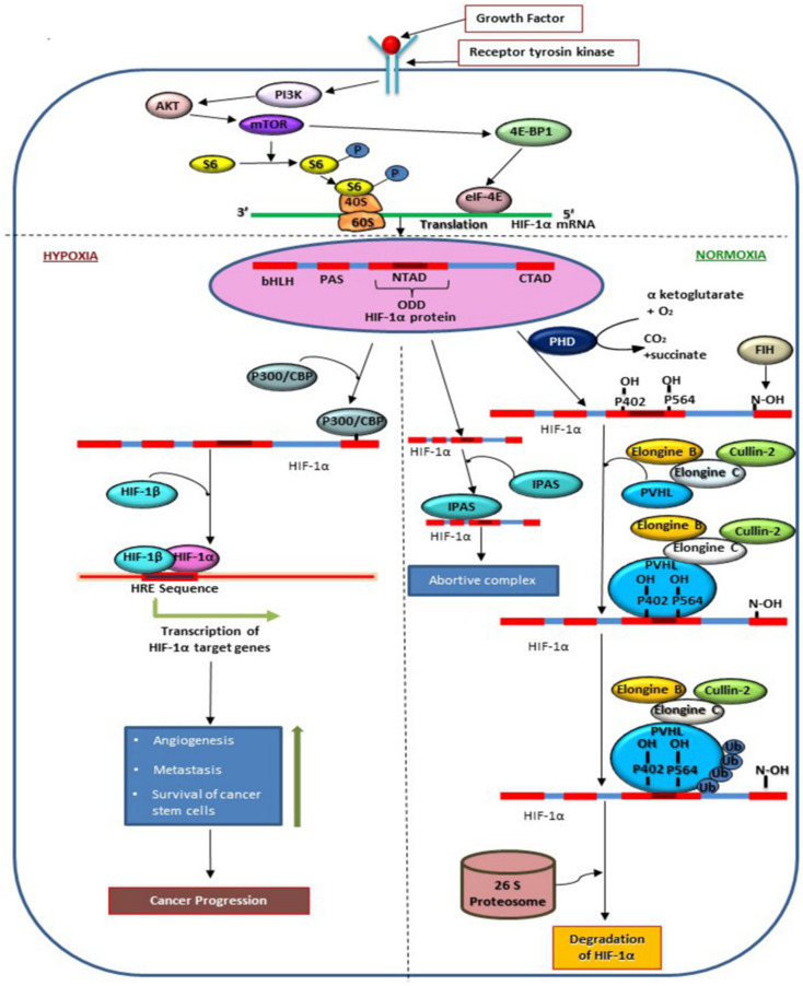
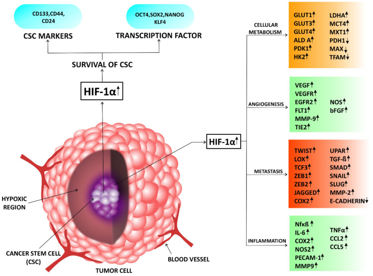
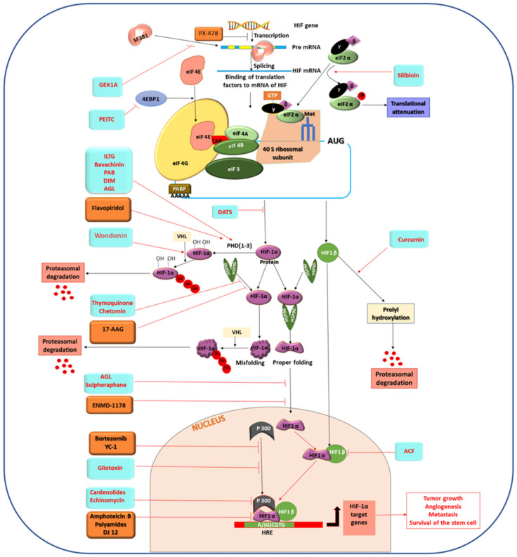
Advancement in novel target detection using improved molecular cancer biology has opened up new avenues for promising anti-cancer drug development. In the past two decades, the mechanism of tumor hypoxia has become more understandable with the discovery of hypoxia-inducible factor-1α (HIF-1α). It is a major transcriptional regulator that coordinates the activity of various transcription factors and their downstream molecules involved in tumorigenesis. HIF-1α not only plays a crucial role in the adaptation of tumor cells to hypoxia but also regulates different biological processes, including cell proliferation, survival, cellular metabolism, angiogenesis, metastasis, cancer stem cell maintenance, and propagation. Therefore, HIF-1α overexpression is strongly associated with poor prognosis in patients with different solid cancers. Hence, pharmacological targeting of HIF-1α has been considered to be a novel cancer therapeutic strategy in recent years. In this review, we provide brief descriptions of natural and synthetic compounds as HIF-1α inhibitors that have the potential to accelerate anticancer drug discovery. This review also introduces the mode of action of these compounds for a better understanding of the chemical leads, which could be useful as cancer therapeutics in the future.

Keywords: HIF-1α; angiogenesis; cancer stem cells; hypoxia; metastasis; natural compounds; synthetic drugs.


Conflict of interest statement

The authors declare no conflict of interest.

Figures

Figure 1
Figure 1
Molecular mechanism of regulation of HIF-1α in hypoxic and normoxic conditions. The figure represents the induction of HIF-1α translation via PI3K/AKT/mTOR pathway. On binding to growth factors at receptor tyrosin kinase PI3K becomes activated which further induces AKT and mTOR pathway activation followed by phosphorylation of S6. HIF-1α synthesis is induced by eIF-4E which binds to HIF-1 α upon activation by 4E-BP1 which is a downstream signaling molecule of mTOR. In the presence of oxygen, Pro-402, Pro-564 in ODD and Asn-803 in CTAD are hydroxylated by PHD and FIH. As represented in the figure, hydroxylation at Asn-803 prevents binding of P300/CBP to HIF-1 α in normoxic conditions, whereas, hydroxylation at Proline residues allow VHL- elongine-C-elongine-B-Cullin-2 complex to bind at ODD of HIF-1α followed ubiquitination of HIF-1α via 26 proteasome. Expression of HIF-1α is also regulated by IPAS, a variant of HIF-3 which binds with HIF-1α to form an abortive complex. In hypoxic conditions, P300/CBP binds at CTAD which prevents degradation of HIF-1α. HIF-1α enters nucleus and forms active transcription factor by binding with HIF-1β in order to transcribe genes for angiogenesis, metastasis, and survival of cancer stem cells in tumor tissue.
Figure 2
Figure 2
Representation of target genes of HIF-1α involved in tumor progression: Figure represents tumor tissue with hypoxic region at its core. Cancer stem cell (CSC) population resides at the core region. In hypoxic tumor tissue, HIF-1α helps in regulation of genes involved in cellular metabolism [41,42,43,44,45,46,47,48,49,50,51,52,53,54,55,56,57,58,59,60,61,62,63,64,65,66], angiogenesis [67,68,69,70,71,72,73,74,75,76,77,78,79,80,81,82,83,84,85,86], metastasis [87,88,89,90,91,92,93,94,95,96,97,98,99,100,101,102], CSC propagation & maintenance [103,104,105,106,107,108,109,110,111] and cancer inflammation [112,113,114,115,116,117,118,119,120,121,122] which are listed in the figure. Upward arrows (↑) indicate increased expression of the protein and Downward arrows (↓) indicate decreased expression of the protein.
Figure 3
Figure 3
Molecular mechanism of different natural and synthetic compounds targeting HIF-1α and its pathways. The figure represents sky blue and brown boxes that symbolize natural and synthetic compounds, respectively. The illustration denotes GEX1A-mediated inhibition of HIF-1α mRNA splicing by inactivating spliceosome core protein SF3B1. The figure also shows translational attenuates of HIF-1α by silibinin via phosphorylation of α subunit of eIF2. ILTG, Bavachinin, PAB, DIM, AGL, and Flavopiridol-induced prolyl hydroxylation mediated proteasomal degradation of HIF-1α protein is also represented in this figure. Down-regulation of the activity of HIF-1α through proteasomal degradation of its interacting subunit αβ by Curcumin is also demonstrated here. This figure shows the mode of action of Chetomin and Thymoquinone by blocking HSP 90 mediated folding of HIF-1α protein which causes proteasomal degradation of it. Inhibition of nuclear localization of HIF-1α by AGL and Sulforaphane is shown in the figure. Suppression of HIF-1α and HIF-1β heterodimer formation by ACF is also represented. The figure denotes Bortezomib and Gliotoxin dependent inhibition of the interaction between p300 and HIF-1α. Cardenolides, Echinomycin, Amphotericin B, polyamides and DJ12 suppress the binding of HIF-1α/p300 complex to HRE, which are also represented in this figure.

References

    1. Müller M., Padberg W., Schindler E., Sticher J., Osmer C., Friemann S., Hempelmann G. Renocortical tissue oxygen pressure measurements in patients undergoing living donor kidney transplantation. Anesth. Analg. 1998;87:474–476. doi: 10.1213/00000539-199808000-00045. - DOI - PubMed
    1. Dings J., Meixensberger J., Jäger A., Roosen K. Clinical experience with 118 brain tissue oxygen partial pressure catheter probes. Neurosurgery. 1998;43:1082–1095. doi: 10.1097/00006123-199811000-00045. - DOI - PubMed
    1. Vaupel P., Thews O., Hoeckel M. Treatment resistance of solid tumors: Role of hypoxia and anemia. Med. Oncol. Northwood Lond. Engl. 2001;18:243–259. doi: 10.1385/MO:18:4:243. - DOI - PubMed
    1. Vaupel P. Tumor microenvironmental physiology and its implications for radiation oncology. Semin. Radiat. Oncol. 2004;14:198–206. doi: 10.1016/j.semradonc.2004.04.008. - DOI - PubMed
    1. Aggarwal V., Tuli H.S., Varol A., Thakral F., Yerer M.B., Sak K., Varol M., Jain A., Khan M.A., Sethi G. Role of reactive oxygen species in cancer progression: Molecular mechanisms and recent advancements. Biomolecules. 2019;9:735. doi: 10.3390/biom9110735. - DOI - PMC - PubMed

MeSH terms

Substances