. 2022 Aug 16;14(16):3359.
doi: 10.3390/nu14163359.

Serum Vitamin D Levels Explored in the Latvian Cohort of Patients with Basal Cell Carcinoma Linked to the Sonic Hedgehog and Vitamin D Binding Protein Cutaneous Tissue Indices

Affiliations

Serum Vitamin D Levels Explored in the Latvian Cohort of Patients with Basal Cell Carcinoma Linked to the Sonic Hedgehog and Vitamin D Binding Protein Cutaneous Tissue Indices

Jeļena Moisejenko-Goluboviča et al. Nutrients. .

Abstract

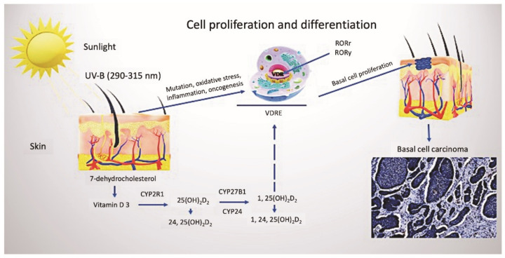
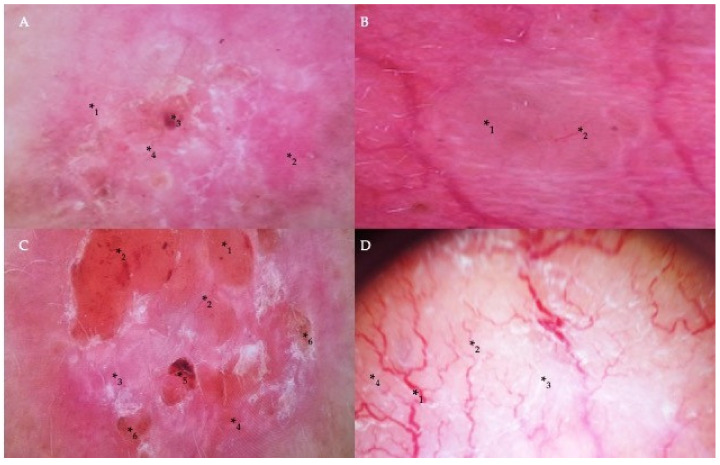
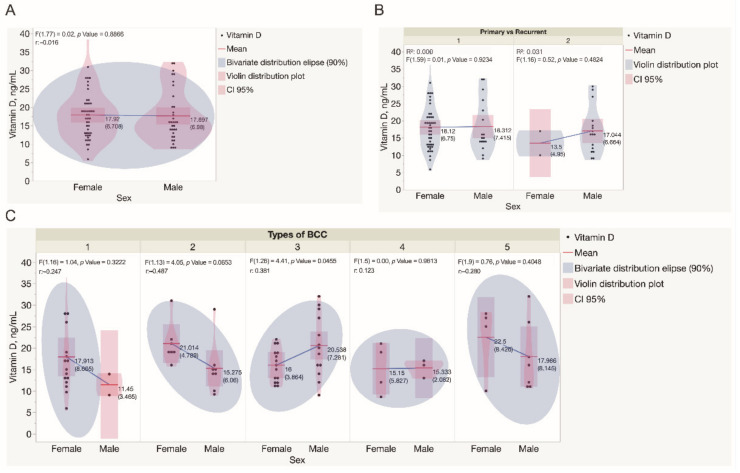
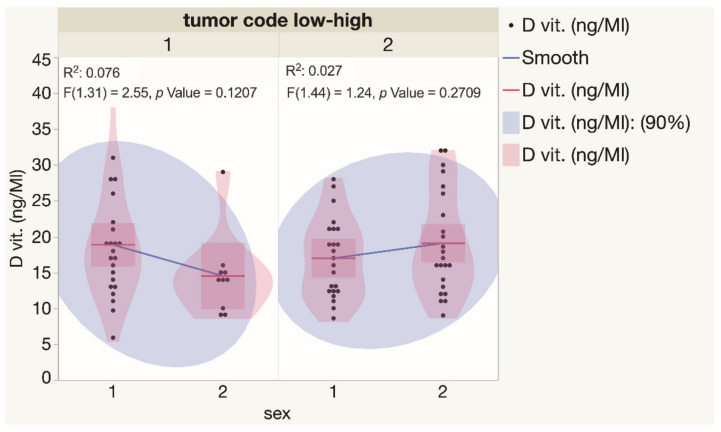
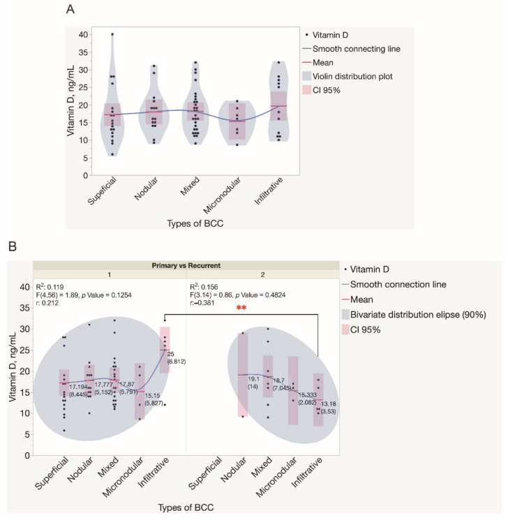
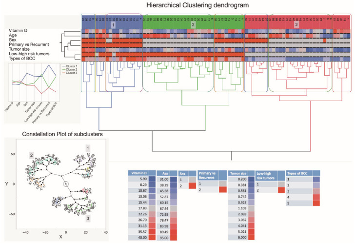
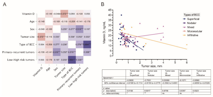
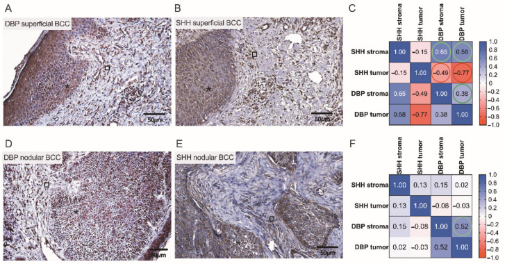
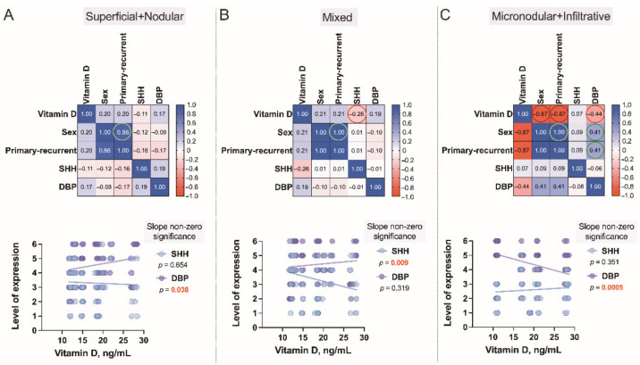
Ultraviolet radiation is known as one of the major contributors to skin malignancies, including basal cell carcinoma (BCC), which is the most common type of skin cancer. It is a heterogeneous tumor, which presents with various types that are stratified into low- and high-risk tumors. Sunlight is important for overall health and vitamin D synthesis in the skin, whereas deviations from the optimal level of vitamin D are shown to be associated with the risk of the development of BCC. The accumulating evidence suggests the ability of vitamin D to antagonize the Sonic Hedgehog (SHH) signaling, the key tumor pathway, and play a protective role in the development of BCC. Additionally, a vitamin D binding protein (DBP) is shown to be implicated in the complex regulation of vitamin D. Here, we aimed to explore serum vitamin D in patients with different primary and recurrent BCC of the head and neck and investigate cutaneous DBP and SHH indices, confirmed immunohistochemically in these subjects. According to the results, 94.9% of the Latvian cohort of BCC patients were found to be deficient in vitamin D. No significant differences in serum vitamin D levels were found between genders, primary and recurrent tumors, and different types of BCC. Serum vitamin D was inversely associated with tumor size. Susceptible male individuals with low blood vitamin D levels were recognized at risk of developing aggressive and recurrent BCC confirmed by the use of hierarchical clustering analysis. In smaller tumors with a favorable course, such as superficial and nodular BCC, the association between high DBP and low SHH tissue expression was found, providing supportive evidence of the existence of a link between vitamin D, proteins involved in its metabolism, as exemplified by the DBP and SHH signaling pathway. The assumption of a deficiency in the protective effect of vitamin D in patients with high-risk BCCs was proposed in low DBP and high SHH tissue indices. New extensions to existing knowledge and characterization of the BCC signaling pathways and their cross-talk with vitamin D are warranted when searching for a preferential effect of vitamin D on skin cancer.

Keywords: Sonic Hedgehog; basal cell carcinoma; hierarchical clustering; immunohistochemistry; serum levels of vitamin D; ultraviolet radiation; vitamin D binding protein; vitamin D deficiency.


Conflict of interest statement

The authors declare no conflict of interest.

Figures

Figure 1
Figure 1
Vitamin D metabolism and cutaneous synthesis. Under exposure to UVB rays, the process of vitamin D3 synthesis proceeds in keratinocytes, which contain CYP enzymes necessary for the photochemical conversion of the precursor of vitamin D3, 7-dehydrocholesterol, into its active form (calcitriol 1, 25(OH)2D2). The activities of hydroxyderivatives of vitamin D are mediated by the involvement of the ligand-binding domain of the nuclear receptor, VDR. Furthermore, vitamin D3 is implicated in the regulation of biological functions and gene expression of keratinocytes of both healthy and BCC-affected subjects, mediated by the presence of ROR α and γ nuclear receptors. Abbreviations: UVB, ultraviolet B; VDR, vitamin D receptor; RORα and γ, retinoic acid-related orphan receptors α and γ; BCC, basal cell carcinoma.
Figure 2
Figure 2
Dermoscopic findings in different types of BCC. (A) Milky-pink color structureless areas (*1), pink homogeneous areas (*2), short thin telangiectasia (*3), and erosion (*4) on the central part of the tumor in the case of superficial BCC. (B) A translucent nodule (*1) raised above the skin and arborizing vessels (*2) in nodular BCC. (C) The nodular appearance of a tumor with a border raised above the central part of the lesion (*1), arborizing and short thin telangiectasias (*2), milky-pink (*3) and milky-red color structureless areas (*4), ulceration (*5), and erosion (*6) in the case of micronodular BCC. (D) Arborizing vessels (*1), short thin telangiectasia (*2) on shiny white (*3) and milky-red structureless areas (*4) in the infiltrative and mixed type of BCC.
Figure 3
Figure 3
Assessment of serum vitamin D levels in the study cohort. (A) Assessment of serum vitamin D levels in males and females recruited in the study. Each dot represents a single data point. (B) Assessment of serum vitamin D levels in males and females presented with either primary or recurrent BCC tumors. Each dot represents a single data point. (C) Assessment of serum vitamin D levels in males and females presented with different types of BCC tumors. Each dot represents a single data point. Statistically significant differences in serum vitamin D levels were not observed when different types of BCC diagnosed in both genders were compared, except in mixed BCC tumors, diagnosed in females presented with significantly lower serum vitamin D levels when compared to males (p = 0.0456). Abbreviations: (B) 1—primary BCC tumors; 2—recurrent BCC tumors; (C) 1—superficial BCC tumors; 2—nodular BCC tumors; 3—mixed BCC tumors; 4—micronodular BCC tumors; 5—infiltrative BCC tumors.
Figure 4
Figure 4
Assessment of serum vitamin D levels in males and females presented with low and high risk BCCs. Each dot represents a single data point. Abbreviations: 1—low-risk BCC tumors; 2—high-risk BCC tumors.
Figure 5
Figure 5
Assessment of serum vitamin D levels in patients presented with different types of BCC tumors. (A) Violin distribution plots depict the results of the assessment of serum vitamin D levels in patients presented with different types of BCC tumors. Each dot represents a single data point. (B) The results of the assessment of serum vitamin D levels in patients presented with either primary or recurrent BCC tumors. Each dot represents a single data point. Abbreviations: (B) 1—primary BCC tumors; 2—recurrent BCC tumors. ** p < 0.01.
Figure 6
Figure 6
A dendrogram shows hierarchical clustering—relationships between sets of data. The dendrogram consists of stacked branches (clades) that break down into further smaller branches. At the lowest level, individual elements appear and then they are grouped according to attributes into clusters with fewer and fewer clusters on higher levels. The end of each clade (a leaf) are the data. The sets of data included assessments of serum vitamin D level patterns of 79 subjects presenting with the different types of BCC, information about a patient age and sex, a tumor type (primary vs. recurrent), and a type of BCC, and seen in the right lower part of the Figure. The tumor specimens were divided into five subtypes based on differences in histopathology and dermoscopic imaging. The three major data clusters with 2–3 subclusters each are highlighted in the constellation plot of the dendrogram depicted in the left lower part of the Figure. Abbreviations: sex: 1—females; 2—males; primary vs. recurrent BCC: 1—primary BCC; 2—recurrent BCC; low and high-risk BCC: 1—low risk BCC; 2—high risk BCC; types of BCC: 1—superficial BCC tumors; 2—nodular BCC tumors; 3—mixed BCC tumors; 4—micronodular BCC tumors; 5—infiltrative BCC tumors.
Figure 7
Figure 7
(A) A correlogram of the studied variables. In this plot, correlation coefficients are colored according to the value. Positive correlations are displayed in blue, whereas negative correlations are in red. Color intensity is proportional to the correlation coefficients. The negative association (r = −0.372) between the tumor size and serum vitamin D level is marked by a dark red color. (B) Correlation between the tumor size and serum vitamin D level was observed in different types of BCC. * p < 0.05; ** p < 0.01; *** p < 0.001.
Figure 8
Figure 8
Assessment of cutaneous DBP and SHH expression in low risk BCCs by the use of IHC. Representative images demonstrating cutaneous structures (tumoral (⁎) and stromal (□)) decorated by the anti-DBP (A,D) and anti-SHH (B,E) antibodies and recognized by the presence of brown reaction products found in the BCC samples. Scale bars: 50 μm. Correlograms that highlight associations between the expression of DBP and SHH in superficial (C) and nodular (F) BCCs. A strong negative correlation between SHH and DBP expressions is marked by red color (C).
Figure 9
Figure 9
Assessment of cutaneous DBP and SHH expression in high risk BCCs by the use of IHC. Representative images demonstrating cutaneous structures (tumoral (⁎) and stromal (□)) decorated by the anti-DBP (A,D,G) and anti-SHH (B,E,H) antibodies and recognized by the presence of brown reaction products found in the BCC samples. Scale bars: 50 μm. Correlograms that highlight associations between the expression of DBP and SHH in mixed (C), micronodular (F), and infiltrative (I) BCCs. A negative correlation between SHH and DBP expressions is marked by red color (I).
Figure 10
Figure 10
Correlograms of the studied variables, Superficial+Nodular (A), Mixed (B) and Micronodular+Infiltrative (C). The correlograms depict the complex associations between serum vitamin D level patterns of 79 subjects presenting with the different types of BCC and the cutaneous tissue expression of vitamin D binding protein (DBP) and Sonic Hedgehog (SHH) confirmed by the use of IHC in these individuals. Each dot represents a single data point. The levels of expression are assessed as a sum of the tumoral and stromal BCC tissue indices. In these plots, correlation coefficients are colored according to the value. Positive correlations are displayed in blue, whereas negative correlations are in red. Color intensity is proportional to the correlation coefficients.

References

    1. Situm M., Buljan M., Bulat V., Lugović-Mihić L., Bolanca Z., Simić D. The role of UV radiation in the development of basal cell carcinoma. Coll. Antropol. 2008;32:167–170. - PubMed
    1. Birch-Johansen F., Jensen A., Mortensen L., Olesen A.B., Kjaer S.K. Trends in the incidence of nonmelanoma skin cancer in Denmark 1978–2007: Rapid incidence increase among young Danish women. Int. J. Cancer. 2010;127:2190–2198. doi: 10.1002/ijc.25411. - DOI - PubMed
    1. Tampa M., Georgescu S.R., Mitran C.I., Mitran M.I., Matei C., Scheau C., Constantin C., Neagu M. Recent Advances in Signaling Pathways Comprehension as Carcinogenesis Triggers in Basal Cell Carcinoma. J. Clin. Med. 2020;9:3010. doi: 10.3390/jcm9093010. - DOI - PMC - PubMed
    1. Lee J.W., Ratnakumar K., Hung K.F., Rokunohe D., Kawasumi M. Deciphering UV-induced DNA Damage Responses to Prevent and Treat Skin Cancer. Photochem. Photobiol. 2020;96:478–499. doi: 10.1111/php.13245. - DOI - PMC - PubMed
    1. Harris R.B., Griffith K., Moon T.E. Trends in the incidence of nonmelanoma skin cancers in southeastern Arizona 1985-1996. J. Am. Acad. Dermatol. 2001;45:528–536. doi: 10.1067/mjd.2001.114742. - DOI - PubMed