. 2022 Jul 29;11(8):853.
doi: 10.3390/pathogens11080853.

Interferon Epsilon Signaling Confers Attenuated Zika Replication in Human Vaginal Epithelial Cells

Affiliations

Interferon Epsilon Signaling Confers Attenuated Zika Replication in Human Vaginal Epithelial Cells

James W Mungin Jr et al. Pathogens. .

Abstract

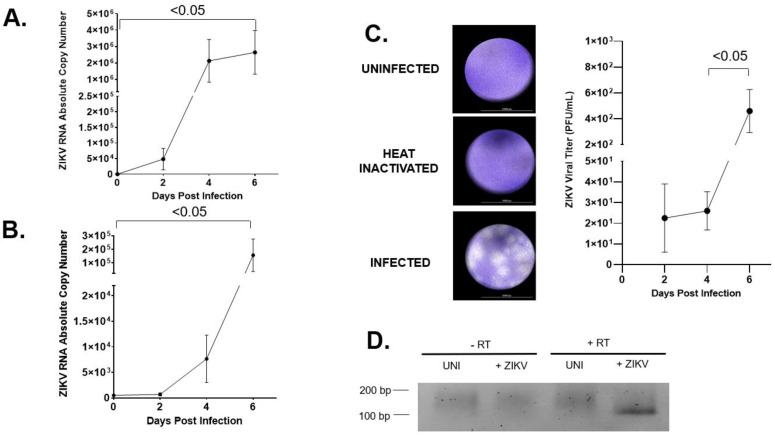
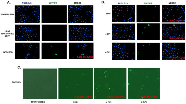
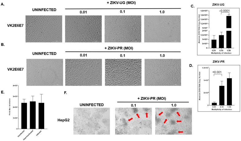
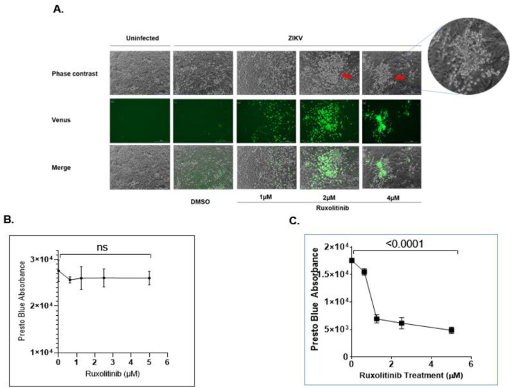
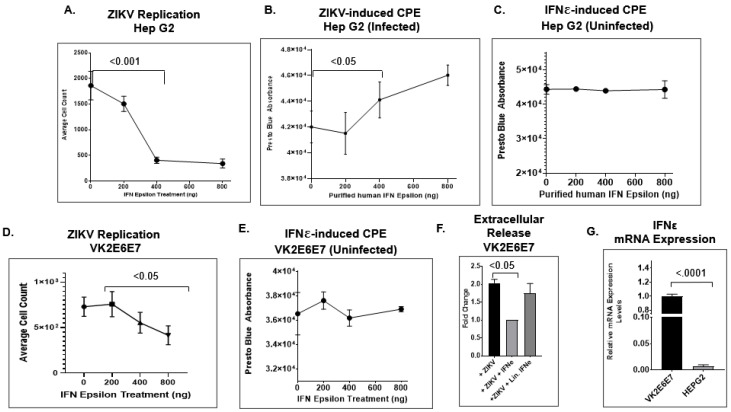
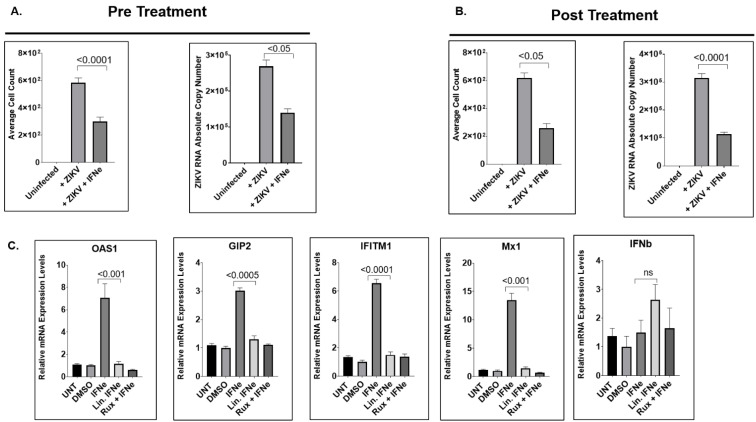
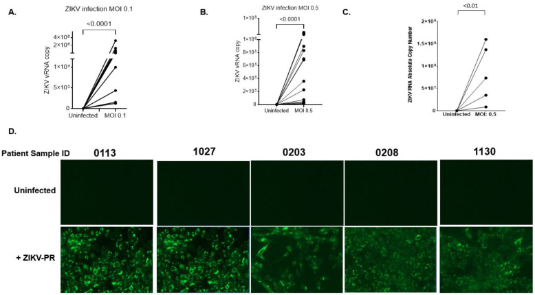
Zika virus (ZIKV) is an emerging flavivirus that causes congenital birth defects and neurological compilations in the human host. Although ZIKV is primarily transmitted through infected mosquitos, recent studies reveal sexual contact as a potential transmission route. In vagina-bearing individuals, the vaginal epithelium constitutes the first line of defense against viruses. However, it is unclear how ZIKV interacts with the vaginal epithelium to initiate ZIKV transmission. In this study, we demonstrate that exposing ZIKV to human vaginal epithelial cells (hVECs) resulted in de novo viral RNA replication, increased envelope viral protein production, and a steady, extracellular release of infectious viral particles. Interestingly, our data show that, despite an increase in viral load, the hVECs did not exhibit significant cytopathology in culture as other cell types typically do. Furthermore, our data reveal that the innate antiviral state of hVECs plays a crucial role in preventing viral cytopathology. For the first time, our data show that interferon epsilon inhibits ZIKV replication. Collectively, our results in this study provide a novel perspective on the viral susceptibility and replication dynamics during ZIKV infection in the human vaginal epithelium. These findings will be instrumental towards developing therapeutic agents aimed at eliminating the pathology caused by the virus.

Keywords: Zika virus; human vaginal epithelial cells; interferon epsilon; primary cervical cell; sexual transmission.


Conflict of interest statement

The authors declare no conflict of interest.

Figures

Figure 1
Figure 1
hVECs support ZIKV replication and viral production. (A) VK2 cell was infected with ZIKV-PR at a low multiplicity infection of 0.01. At days 0, 2, 4, and 6, positive-sense viral RNA were extracted from cell lysate and subjected to qRT-PCR analysis to monitor the dynamics of the vRNA. (B) Extracellular viral release from the culture supernatant of the same infected cells as in (A) was measured using qRT-PCR. (C) PFU viral titer from the supernatant of ZIKV-infected VK2 cells at 2, 4, and 6 days post-infection were quantified and plotted. (D) Negative-sense viral RNA, which is a molecular marker for de novo viral replication, was analyzed from infected cell lysates harvested at day 6 post-infection using RT-PCR. The PCR product of negative-sense vRNA was located at 102 bp. The t test was used to measure p-values.
Figure 2
Figure 2
hVECs support ZIKV replication and viral production. (A) VK2 cells were infected with ZIKV-PR (MOI: 0.5). Uninfected and heat-inactivated ZIKV cells were used as controls. Viral replication was monitored through the presence of the viral envelope (E) protein using an indirect immunofluorescence assay (IFA) with the 4G2 primary antibody and fluorescein isothiocyanate (FITC)-conjugated secondary antibody. DAPI was used to stain the nucleus. (B) VK2 was infected with ZIKV-PR (MOI: 0.5). The time course of viral replication was analyzed using IFA, as mentioned above. (C) VK2 cells were infected with ZIKV-UG (MOI: 0.5). Venus fluorescent protein expression cells were visualized with a BioTek LionHeart FX Automated Microscope. The percentages of cells positive for ZIKV-PR were calculated by dividing the average number of ZIKV-infected cells (GFP signal) by the average number of cells. The calculated percentages are located at the lower right corners of the merged images. The total numbers of VK2E6E7 cells expressing the Venus fluorescent protein through ZIKV-UG were quantified using the BioTek LionHeart FX Automated Microscope.
Figure 3
Figure 3
hVECs do not exhibit significant cytopathic effects following ZIKV exposure. VK2 cells were exposed to ZIKV-UG (A) or ZIKV-PR (B) at different multiplicities of infection (0.01, 0.1, and 1.0) and later examined at day six post-infection. To confirm active ZIKV infection, the supernatant from the same well shown in (A,B) was monitored using qRT-PCR. The viral RNA copy numbers were respectively quantified (C,D). Virus-induced cytotoxicity was measured biochemically using the PrestoBlue assay. The PrestoBlueTM reagent was added to the wells of uninfected, heat-inactivated, and ZIKV-PR-infected VK2E6E7 cells, which were incubated at 37 °C for at least 10 min. The absorbance of each well was quantified using a microplate reader. The relative absorbance was calculated by subtracting the absorbance of the tested wells from the absorbance reads from the culture media alone (E). Data were expressed as the means ± SD of triplicate experiments. To ensure that a viral cytopathic effect occurred, ZIKV-infected VK2 cells were examined alongside ZIKV-infected HEPG2 (human liver epithelial cell line) cells using bright field microscopy. Red arrows indicate CPE formation (F). The t-test was used to measure p-values.
Figure 4
Figure 4
Interferon pathway inhibitor ruxolitinib enhances ZIKV replication and induces cytopathic effects in ZIKV-infected hVECs. (A) Different concentrations of ruxolitinib were used to treat ZIKV-UG-infected VK2 cells. DMSO treatment was used as a control. At day 7 post-infection, images were recorded using a BioTek LionHeart FX Automated Microscope. The red arrows indicate CPE formation. Cell viability for uninfected (B) and infected (C) VK2E6E7 cells treated with ruxolitinib was quantified by subjecting the cultured cells to a PrestoBlue assay. The absorbance of each well was quantified using a microplate reader. The relative absorbance was calculated by subtracting the absorbance of the tested wells from the absorbance reads from the culture media alone. The t-test was used to measure p- values. ns, not significant.
Figure 5
Figure 5
IFNε attenuates ZIKV replication. (A) Hep G2 cells were infected with ZIKV-UG (MOI: 0.25). Different concentrations of human IFNε were used to treat the infected cells. Five days post-infection, the Venus fluorescent protein-expressing cells were visualized using a BioTek LionHeart FX Automated Microscope. The total number of Venus fluorescent protein-expressing cells was plotted with the concentration of IFNε. Cytopathic effects from both ZIKV-infected (B) and uninfected (C) HEPG2 cells were measured using the PrestoBlue assay, in which the relative absorbance of live, viable cells was quantified using a microplate reader. (D) VK2E6E7 cells were infected with the ZIKV-UG strain alongside different concentrations of purified human IFNε. The average cell count was calculated by quantifying the Venus fluorescent protein-expressing cells using a BioTek LionHeart FX Automated Microscope. (E) The CPEs of uninfected VK2E6E7 treated with different concentrations of IFNε were measured with PrestoBlue and quantified using a microplate reader. (F) Extracellular viral release was measured by collecting the culture supernatant from experimental samples and quantifying the absolute viral RNA copy number using qPCR analysis. (G) Using qPCR analysis, the expression profiles of IFNε for the HEP G2 and VK2E6E7 cell lines were measured, and 18 s rRNA was used as a normalizing control. An unpaired t-test was used to measure p-values.
Figure 6
Figure 6
IFNε treatment restricts ZIKV replication via induction of Type I Interferon stimulated genes. (A) Effect of pretreatment with IFN-ε on ZIKV infection in hVECs. VK2E6E7 cells were stimulated with recombinant IFN-ε for one hour prior to ZIKV infection with the ZIKV-UG strain. Viral replication and extracellular release were measured at five days post-infection. (B) Antiviral effects of IFN-ε treatment after ZIKV infection. VK2E6E7 cells were infected with ZIKV-UG and later treated with IFN-ε after infection. (C) IFN-ε induces type I IFN-stimulated genes (ISGs). VK2E6E7 cells were treated with IFN-ε for six hours, and gene expression of ISGs was measured by real-time qPCR analysis. Relative mRNA expression levels (as fold change relative to DMSO control) for each gene of interest were calculated using the ΔΔCT method, as described in the Materials and Methods Section. The normalizing control used for the qPCR was the housekeeping GUSB gene. An unpaired t-test was used to analyze the p-values for the viral replication and induction of ISGs.
Figure 7
Figure 7
Primary human cervical cells are susceptible to ZIKV infection. (A,B) Primary human cervical cells from 15 donors were infected with ZIKV-PR (MOI: 0.1 or 0.5). After infection, the cells were extensively washed and culture for four days. Four days post-infection, the culture supernatant was subjected to qRT-PCR to measure viral RNA copy numbers. (C) To determine infectivity, the cultured supernatant collected from the primary cells infected with ZIKV-PR (n = 5) was used as inoculum to infect Vero cells. The absolute RNA copy number was determined by subjecting the supernatant collected from Vero cells to qPCR analysis. (D) In addition to qPCR analysis, active viral production was examined by performing IFA on Vero cells infected by the inoculum. The Wilcoxon matched-pairs signed-rank test was used to analyze the significance of viral replication.

Similar articles

Cited by

References

    1. Song B.H., Yun S.I., Woolley M., Lee Y.M. Zika virus: History, epidemiology, transmission, and clinical presentation. J. Neuroimmunol. 2017;308:50–64. doi: 10.1016/j.jneuroim.2017.03.001. - DOI - PubMed
    1. Sharma V., Sharma M., Dhull D., Sharma Y., Kaushik S., Kaushik S. Zika virus: An emerging challenge to public health worldwide. Can. J. Microbiol. 2020;66:87–98. doi: 10.1139/cjm-2019-0331. - DOI - PubMed
    1. Runge-Ranzinger S., Morrison A.C., Manrique-Saide P., Horstick O. Zika transmission patterns: A meta-review. Trop. Med. Int. Health. 2019;24:523–529. doi: 10.1111/tmi.13216. - DOI - PubMed
    1. Petersen L.R., Jamieson D.J., Powers A.M., Honein M.A. Zika Virus. N. Engl. J. Med. 2016;374:1552–1563. doi: 10.1056/NEJMra1602113. - DOI - PubMed
    1. Musso D., Guble D.J. Zika Virus. Clin. Microbiol. Rev. 2016;29:487–524. doi: 10.1128/CMR.00072-15. - DOI - PMC - PubMed

LinkOut - more resources