

A Recombinant Vaccine-like Strain of Lumpy Skin Disease Virus Causes Low-Level Infection of Cattle through Virus-Inoculated Feed
- PMID: 36015041
- PMCID: PMC9414542
- DOI: 10.3390/pathogens11080920
A Recombinant Vaccine-like Strain of Lumpy Skin Disease Virus Causes Low-Level Infection of Cattle through Virus-Inoculated Feed
Abstract
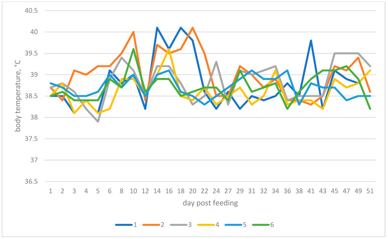
Since 1989, lumpy skin disease of cattle (LSD) has spread out of Africa via the Middle East northwards and eastwards into Russia, the Far East and South-East Asia. It is now threatening to become a worldwide pandemic, with Australia possibly next in its path. One of the research gaps on the disease concerns its main mode of transmission, most likely via flying insect vectors such as biting flies or mosquitoes. Direct or indirect contact transmission is possible, but appears to be an inefficient route, although there is evidence to support the direct contact route for the newly detected recombinant strains first isolated in Russia. In this study, we used experimental bulls and fed them via virus-inoculated feed to evaluate the indirect contact route. To provide deeper insights, we ran two parallel experiments using the same design to discover differences that involved classical field strain Dagestan/2015 LSDV and recombinant vaccine-like Saratov/2017. Following the attempted indirect contact transmission of the virus from the inoculated feed via the alimentary canal, all bulls in the Dagestan/2015 group remained healthy and did not seroconvert by the end of the experiment, whereas for those in the Saratov/2017 recombinant virus group, of the five bulls fed on virus-inoculated feed, three remained clinically healthy, while two displayed evidence of a mild infection. These results provide support for recombinant virus transmission via the alimentary canal. In addition, of particular note, the negative control in-contact bull in this group exhibited a biphasic fever at days 10 and 20, developed lesions from day 13 onwards, and seroconverted by day 31. Two explanations are feasible here: one is the in-contact animal was somehow able to feed on some of the virus-inoculated bread left over from adjacent animals, but in the case here of the individual troughs being used, that was not likely; the other is the virus was transmitted from the virus-fed animals via an airborne route. Across the infected animals, the virus was detectable in blood from days 18 to 29 and in nasal discharge from days 20 to 42. Post-mortem and histological examinations were also indicative of LSDV infection, supporting further evidence for rapid, in F transmission of this virus. This is the first report of recombinant LSDV strain transmitting via the alimentary mode.
Keywords: lumpy skin disease; recombinant; transmission; virus.
Conflict of interest statement
The authors report no conflict of interest.
Figures
References
-
- World Organisation for Animal Health (OIE) Terrestrial Animal Health Code. Volume 2 OIE; Paris, France: 2019.
-
- Fauquet C.M., Mayo M.A., Maniloff J., Desselberger U., Ball L.A. Virus Taxonomy. Academic Press; Cambridge, MA, USA: 2005. The Double Stranded DNA Viruses; pp. 37–276. - DOI
-
- Davies F.G. Lumpy skin disease of cattle: A growing problem in Africa and the Near East. World Anim. Rev. 1991;68:37–42.
Grants and funding
LinkOut - more resources
Full Text Sources
Miscellaneous
