. 2022 Jul 31;15(8):954.
doi: 10.3390/ph15080954.

Molecular Targets of Pinocembrin Underlying Its Regenerative Activities in Human Keratinocytes

Affiliations

Molecular Targets of Pinocembrin Underlying Its Regenerative Activities in Human Keratinocytes

Jirapak Ruttanapattanakul et al. Pharmaceuticals (Basel). .

Abstract

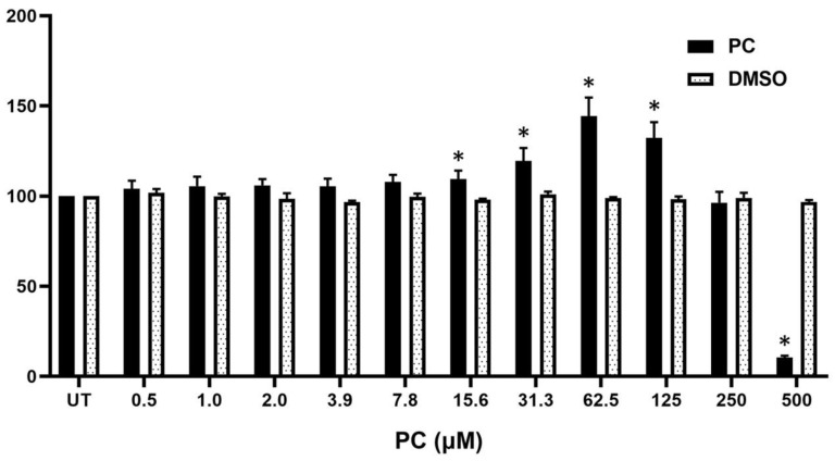
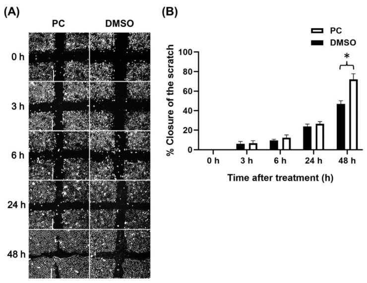
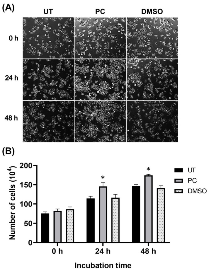
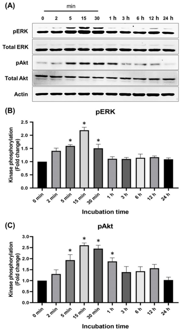
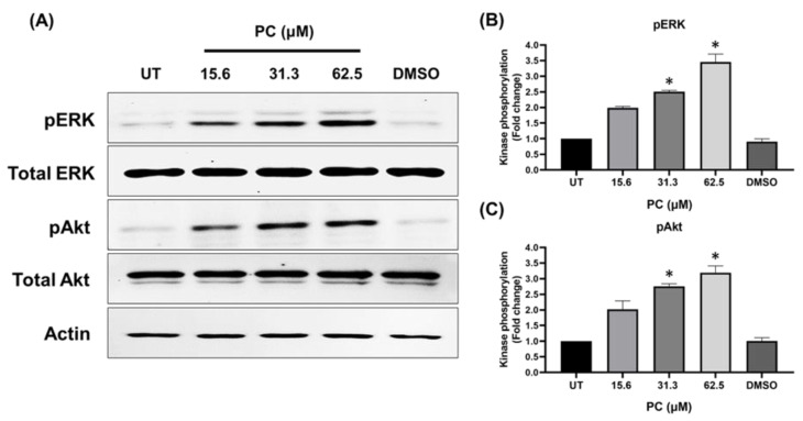
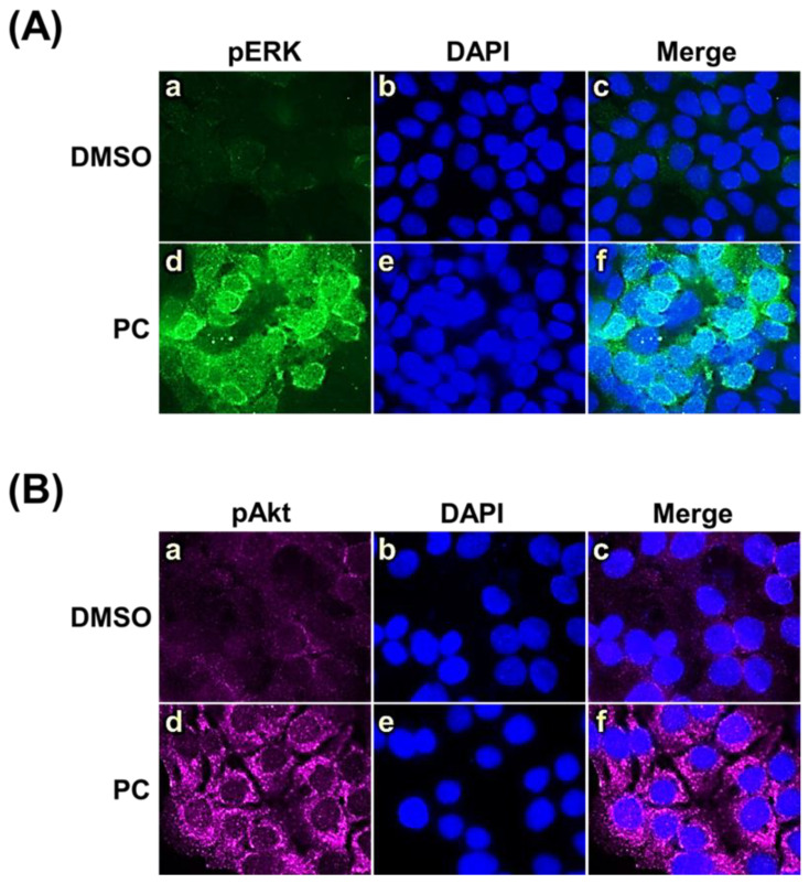
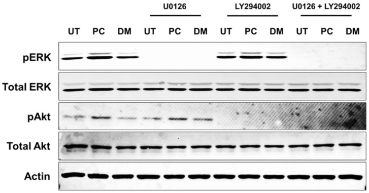
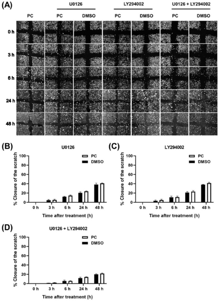
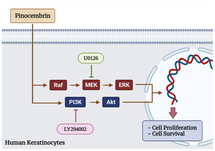
Pinocembrin is one of the well-known compounds in the group of flavonoids. The pharmacological activities of pinocembrin in association with wound-healing activities have been reported. However, its effects on the aspect of cellular interaction underlying growth and survival are still unidentified in human keratinocytes. Our previous study reported that Boesenbergia rotunda potently stimulated survival and proliferation of a human keratinocyte cell line (HaCaT). On the basis that pinocembrin is revealed to be one of the major constituents of this plant, we aimed to define the survival- and proliferation-enhancing effects of this compound at the cellular level. Results from the current study confirmed that pinocembrin induced an increase in HaCaT cell number. At the signaling perspective, we identified that pinocembrin significantly triggered ERK1/2 and Akt activation. The stimulating effects of pinocembrin were clearly inhibited by MEK and PI3K inhibitors authenticating that proliferation- and survival-promoting activities of pinocembrin were mainly acted on these two signaling cascades. Altogether, we successfully identified that pinocembrin functions to induce keratinocyte proliferation and survival, at least by provoking MAPK and PI3K pathways. Our study encourages the fact that pinocembrin is one of the interesting natural flavonoid compounds to be developed as a wound closure-promoting agent.

Keywords: flavonoids; keratinocyte; pinocembrin; regenerative medicine; wound healing.


Conflict of interest statement

The authors declare no conflict of interest.

Figures

Figure 1
Figure 1
Cytotoxicity effects of pinocembrin on HaCaT cells determined by MTT assay. Human keratinocytes treated with different doses of pinocembrin for 48 h in serum-free media. Data were shown as percent cell viability/proliferation with mean ± SD. * p < 0.05 as compared with the un-treated group (UT).
Figure 2
Figure 2
Effects of pinocembrin on accelerating closure of scratch wound of human keratinocyte monolayers. (A) Phase-contrast microscopy (10× magnification) of scratch wound-healing assay at various captured times (0 h, 3 h, 6 h, 24 h, and 48 h) in HaCaT treated with 62.5 μM pinocembrin; (B) Percent closure of the scratch wounded areas of human keratinocyte monolayer over the course of 48 h. Data were analyzed from three individual replicates and presented as mean ± SD. * p < 0.05 as compared with the DMSO control.
Figure 3
Figure 3
Effects of pinocembrin on human keratinocyte proliferation. (A) Phase-contrast observation of HaCaT cells treated with pinocembrin at 62.5 μM at 0, 24, and 48 h (10× magnification) compared with the untreated group and the DMSO control group; (B) Direct cell counting for number of human keratinocytes treated with pinocembrin at 62.5 μM and at 0, 24, and 48 h. Data from three experiments were analyzed and presented as mean ± SD. * p < 0.05 (compared to the untreated group, UT).
Figure 4
Figure 4
Effects of pinocembrin on MAPK and PI3K/Akt signaling pathways. (A) The activation status evaluated by the level of phosphorylation of ERK1/2 and Akt in the lysates of cells treated with 62.5 μM of pinocembrin at various time points; (B) Densitometric analysis of phosphorylated ERK1/2 in cells treated with pinocembrin at each time point; (C) Densitometric analysis of phosphorylated Akt in cells treated with pinocembrin at each time point. The total protein expression of each kinase was detected and used for normalization. Data from three experiments were analyzed and presented as mean ± SD. * p < 0.05 (compared to the 0 min group).
Figure 5
Figure 5
Concentration-dependent effects of pinocembrin on ERK1/2 and Akt activation. (A) Western blotting for ERK1/2 and Akt phosphorylation in HaCaT cells incubated with pinocembrin at 15.6, 31.3, or 62.5 μM for 15 min; (B) Densitometric analysis of phosphorylated ERK1/2 in lysates of cells treated with varied concentrations of pinocembrin; (C) Densitometric analysis of phosphorylated Akt in lysates of cells treated with varied concentrations of pinocembrin. Data from three experiments were analyzed and presented as mean ± SD. * p < 0.05 (compared to the control group, UT).
Figure 6
Figure 6
Immunofluorescence study determining ERK1/2 and Akt activation in cells treated with pinocembrin (62.5 μM). (A) Phosphorylated ERK1/2 (pERK1/2) (green); (B) Phosphorylated Akt (pAkt) (green). HaCaT cells were counterstained with DAPI to detect the nuclei (blue). Pictures were captured by a fluorescent microscope at 100× magnification.
Figure 7
Figure 7
Western blot analysis detecting pERK1/2 and pAkt after incubation with pinocembrin at various concentrations (15.6, 31.3, and 62.5 μM) for 15 min in combination with U0126 and LY294002 which are specific inhibitors of MAPK and PI3K signaling. Data are obtained from three individual experiments.
Figure 8
Figure 8
Scratch wound healing assay for investigating the monolayer wound closure accelerating effects of pinocembrin (62.5 μM) in the presence of U0126, LY294002, or combination of both U0126 and LY294002. (A) Phase-contrast microscopy (10× magnification) of scratch wound-healing assay with U0126, LY294002, or combination of U0126 and LY294002 over the course of 48 h. Percent closure of the scratch-wounded areas of human keratinocyte monolayer over the course of 48 h in the presence of (B) U0126, (C) LY294002, or (D) combination of both U0126 and LY294002. Data from three experiments were analyzed and presented as mean ± standard deviation.
Figure 9
Figure 9
Schematic figure illustrating that pinocembrin regulates human keratinocyte proliferation and survival. Pinocembrin increases the proliferation and survival of keratinocytes through both MAPK signal transduction pathway and PI3K/Akt signaling pathway.

Similar articles

Cited by

References

    1. Kasuya A., Tokura Y. Attempts to accelerate wound healing. J. Dermatol. Sci. 2014;76:169–172. doi: 10.1016/j.jdermsci.2014.11.001. - DOI - PubMed
    1. Pastar I., Stojadinovic O., Yin N.C., Ramirez H., Nusbaum A.G., Sawaya A., Patel S.B., Khalid L., Isseroff R.R., Tomic-Canic M. Epithelialization in Wound Healing: A Comprehensive Review. Adv. Wound Care. 2014;3:445–464. doi: 10.1089/wound.2013.0473. - DOI - PMC - PubMed
    1. Kirsner R.S., Eaglstein W.H. The wound healing process. Dermatol. Clin. 1993;11:629–640. doi: 10.1016/S0733-8635(18)30216-X. - DOI - PubMed
    1. Shi C., Wang C., Liu H., Li Q., Li R., Zhang Y., Liu Y., Shao Y., Wang J. Selection of Appropriate Wound Dressing for Various Wounds. Front. Bioeng. Biotechnol. 2020;8:182. doi: 10.3389/fbioe.2020.00182. - DOI - PMC - PubMed
    1. Han G., Ceilley R. Chronic Wound Healing: A Review of Current Management and Treatments. Adv. Ther. 2017;34:599–610. doi: 10.1007/s12325-017-0478-y. - DOI - PMC - PubMed

LinkOut - more resources