Review
. 2022 Jul 29;14(8):1578.
doi: 10.3390/pharmaceutics14081578.

Progress in 3D Bioprinting Technology for Osteochondral Regeneration

Affiliations
Review

Progress in 3D Bioprinting Technology for Osteochondral Regeneration

Markel Lafuente-Merchan et al. Pharmaceutics. .

Abstract

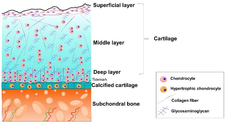
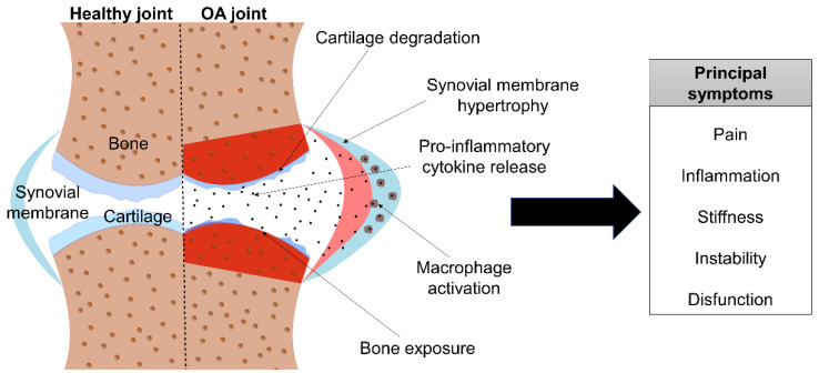
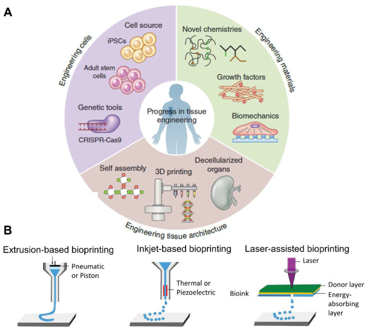
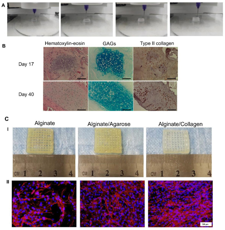
Osteochondral injuries can lead to osteoarthritis (OA). OA is characterized by the progressive degradation of the cartilage tissue together with bone tissue turnover. Consequently, joint pain, inflammation, and stiffness are common, with joint immobility and dysfunction being the most severe symptoms. The increase in the age of the population, along with the increase in risk factors such as obesity, has led OA to the forefront of disabling diseases. In addition, it not only has an increasing prevalence, but is also an economic burden for health systems. Current treatments are focused on relieving pain and inflammation, but they become ineffective as the disease progresses. Therefore, new therapeutic approaches, such as tissue engineering and 3D bioprinting, have emerged. In this review, the advantages of using 3D bioprinting techniques for osteochondral regeneration are described. Furthermore, the biomaterials, cell types, and active molecules that are commonly used for these purposes are indicated. Finally, the most recent promising results for the regeneration of cartilage, bone, and/or the osteochondral unit through 3D bioprinting technologies are considered, as this could be a feasible therapeutic approach to the treatment of OA.

Keywords: 3D bioprinting; bone; cartilage; osteoarthritis; regenerative medicine; tissue engineering.


Conflict of interest statement

The authors declare no conflict of interest. The company had no role in the design of the study; in the collection, analyses, or interpretation of data; in the writing of the manuscript, and in the decision to publish the results.

Figures

Figure 1
Figure 1
The economic cost of knee and hip OA in Spain. Data from [14].
Figure 2
Figure 2
Schematic organization of osteochondral tissue.
Figure 3
Figure 3
Schematic image of OA’s pathology and symptoms.
Figure 4
Figure 4
(A) Diagram of the elements used in tissue engineering. Adapted from [46]. (B) Scheme of different bioprinting methods. Adapted from [47].
Figure 5
Figure 5
Collagen-based scaffolds: (A). Extrusion-based bioprinting of a 4% collagen scaffold. (B). Cartilage ECM evaluation after in vivo implantation. At day 40, GAG accumulation and type II collagen production were increased. Scale bar = 100 µm. Adapted from [57]. (C(I)). Macroscopic images of alginate, alginate–agarose, and alginate–collagen scaffolds. (C(II)). Rhodamine–phalloidin/Hoechst 33,258 staining after 14 days of bioprinting. Scale bar = 100 µm. Adapted from [58].
Figure 6
Figure 6
Alginate/dECM-based scaffolds: (A). Cell viability, histology, and immunostaining on days 0 and 21 showed good cell viability and high GAG and collagen production within 21 days. (B) Alginate/dECM 3D bioprinting and PCL 3D printing combination. (I) Representative image of the hybrid scaffold. (II) Mechanical properties are enhanced with PCL reinforcement. Adapted from [60].
Figure 7
Figure 7
Silk fibroin (SF)-based scaffolds. Extrusion bioprinting process of SF + gelatin bio-ink, obtaining porous scaffolds. Scale bar = 200 µm. Adapted from [64].
Figure 8
Figure 8
In situ bioprinting techniques: (A) In situ crosslinking technique consisting of exposing the bio-ink to visible light just after being extruded. Adapted from [66]. (B) “Biopen”—extrusion-based handled bioprinting technique based on a coaxial system. Adapted from [67].
Figure 9
Figure 9
GelMA-based scaffold: (A) Representative bright-field and fluorescence images of hybrid scaffolds composed of PCL and GelMA. Scale bar = 2 mm. Adapted from [68]. (B) Schematic image of extrusion-based bioprinting and electrowriting techniques that improved scaffold mechanical properties * = p < 0.05. Adapted from [69].
Figure 10
Figure 10
Layered scaffolds: (A) Schematic image of the manufacture of zonally stratified articular cartilage. Adapted from [71]. (B). Histological images of GAGs (safranin-O, top), collagen type II (middle), and collagen type I (bottom) matrix of APCs and MSCs in GelMA/gellan gum/HAMA (GGH) bioprinted scaffolds at day 42. Scale bar = 100 μm. Adapted from [72].
Figure 11
Figure 11
Schematic representation of the bioprinting process with co-printing of PCL and the bioprinting of the bio-ink composed of alginate, MSCs, and nHAP-pDNA complexes. Adapted from [79].
Figure 12
Figure 12
Graphene oxide scaffolds: (A) Optical images of the top view of the printed scaffolds, indicating better printability when GO increases from 0.05 mg/mL to 1 mg/mL. Scale bars = 300 μm. Adapted from [83]. (B) Cell viability in the 3D-bioprinted GO scaffolds at days 1, 7, and 42. Living cells are depicted in green, and dead cells are in red. Scale bar = 50 μm. Adapted from [84].
Figure 13
Figure 13
Multi-tool bioprinting procedure: (A) Schematic images of PCL printing and GelMA and pluronic bioprinting to create the bone region. (B) Macroscopic images of bioprinted scaffold. Live/dead analysis of MSC-laden GelMA bio-ink including microchannels after washing out pluronic. Scale bars = 0.5 mm and 3 mm. (C) Inkjet bioprinting procedure to obtain the cartilage part. Adapted from [88].

References

    1. Xu J., Ji J., Jiao J., Zheng L., Hong Q., Tang H., Zhang S., Qu X., Yue B. 3D Printing for Bone-Cartilage Interface Regeneration. Front. Bioeng. Biotechnol. 2022;10:828921. doi: 10.3389/fbioe.2022.828921. - DOI - PMC - PubMed
    1. Poveda-Roda R., Bagán J.V., Sanchis J., Margaix M. Pseudotumors and Tumors of the Temporomandibular Joint. A Review. Med. Oral Patol. Oral Cirugía Bucal. 2013;18:e392–e402. doi: 10.4317/medoral.18799. - DOI - PMC - PubMed
    1. García-Arias M., Balsa A., Mola E.M. Septic Arthritis. Best practice & research. Clin. Rheumatol. 2011;25:407–421. - PubMed
    1. Chau M.M., Klimstra M.A., Wise K.L., Ellermann J.M., Tóth F., Carlson C.S., Nelson B.J., Tompkins M.A. Osteochondritis Dissecans. J. Bone Jt. Surg. 2021;103:1132. doi: 10.2106/JBJS.20.01399. - DOI - PMC - PubMed
    1. Hawker G.A. OA Serious Disease. Clin. Exp. Rheumatol. 2019;37:3. - PubMed

LinkOut - more resources