. 2022 Aug 2;14(8):1611.
doi: 10.3390/pharmaceutics14081611.

Nanostructured Lipid Carriers Loaded with Dexamethasone Prevent Inflammatory Responses in Primary Non-Parenchymal Liver Cells

Affiliations

Nanostructured Lipid Carriers Loaded with Dexamethasone Prevent Inflammatory Responses in Primary Non-Parenchymal Liver Cells

Carolina Medina-Montano et al. Pharmaceutics. .

Abstract

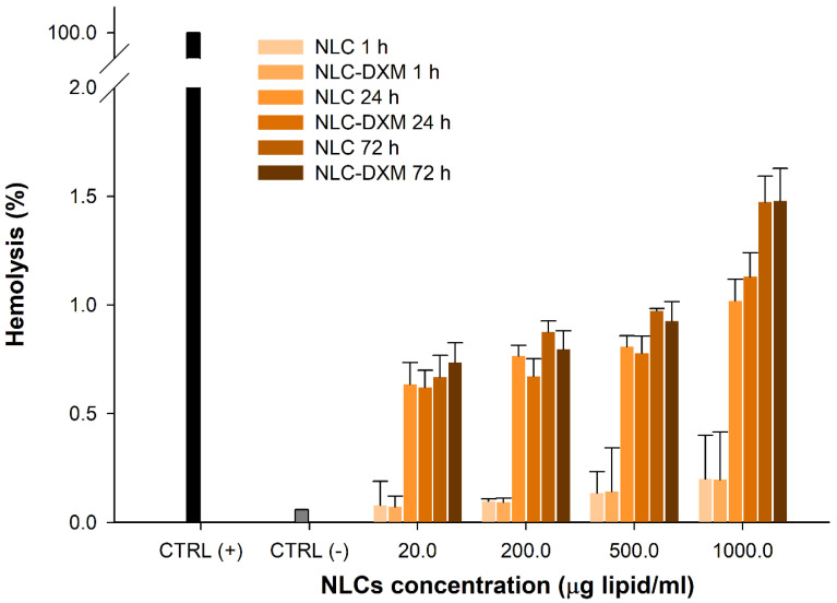
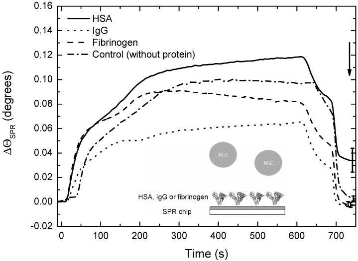
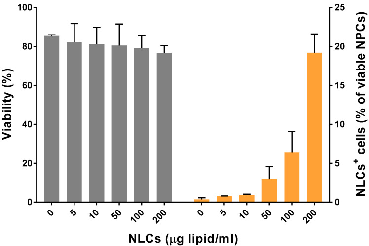
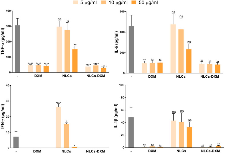
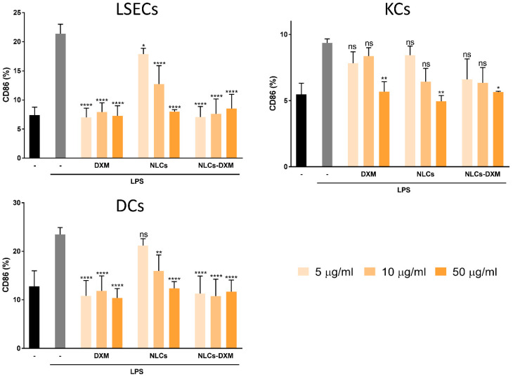
Liver inflammation represents a major clinical problem in a wide range of pathologies. Among the strategies to prevent liver failure, dexamethasone (DXM) has been widely used to suppress inflammatory responses. The use of nanocarriers for encapsulation and sustained release of glucocorticoids to liver cells could provide a solution to prevent severe side effects associated with systemic delivery as the conventional treatment regime. Here we describe a nanostructured lipid carrier developed to efficiently encapsulate and release DXM. This nano-formulation proved to be stable over time, did not interact in vitro with plasma opsonins, and was well tolerated by primary non-parenchymal liver cells (NPCs). Released DXM preserved its pharmacological activity, as evidenced by inducing robust anti-inflammatory responses in NPCs. Taken together, nanostructured lipid carriers may constitute a reliable platform for the delivery of DXM to treat pathologies associated with chronic liver inflammation.

Keywords: autoimmune hepatitis; dexamethasone; drug-controlled release; glucocorticoids; lipid nanoparticles; liver immunology; liver inflammation.


Conflict of interest statement

The authors declare no conflict of interest.

Figures

Figure 1
Figure 1
TEM images of NLCs (F1 to F4 formulations). The freshly prepared formulations (20 mg MM lipid/mL) were diluted with Milli-Q water (1:10) and the contrast was enhanced with phospho-tungstic acid. All images were taken at 30,000× (scale bar = 200 nm).
Figure 2
Figure 2
DXM release inversely correlates with oil content of the NLC matrix. Time-dependent DXM release from the different NLC formulations in 10 mM PBS pH 7.4 and 37 °C, (n = 3).
Figure 3
Figure 3
NLC formulations are stable over time. Stability of NLCs-DXM formulation after 12 months stored at 4 °C. Upper panel: size distribution- Original sample: Mean diameter: 145.0 ± 0.8 nm; PDI: 0.20 ± 0.01; older formulation: size: 150.8 ± 2.5 nm; PDI: 0.24 ± 0.02; lower panel: evolution of the EE measured every 3 months, (n = 3); no significant difference (ns) (Two-way ANOVA, Dunnett’s multiple comparisons test).
Figure 3
Figure 3
NLC formulations are stable over time. Stability of NLCs-DXM formulation after 12 months stored at 4 °C. Upper panel: size distribution- Original sample: Mean diameter: 145.0 ± 0.8 nm; PDI: 0.20 ± 0.01; older formulation: size: 150.8 ± 2.5 nm; PDI: 0.24 ± 0.02; lower panel: evolution of the EE measured every 3 months, (n = 3); no significant difference (ns) (Two-way ANOVA, Dunnett’s multiple comparisons test).
Figure 4
Figure 4
At high concentrations NLCs induce only minimal hemotoxicity. Hemotoxicity of empty and DXM loaded NLCs at different concentrations after 1, 24 and 72 h incubation. Controls: (−) untreated; (+) 1% Triton X-100-lysed erythrocytes, (n = 3).
Figure 5
Figure 5
HSA, but no opsonin interacts with NLCs. SPR studies to determine interactions of DXM-NLCs with serum protein having HSA, IgG and fibrinogen. All sensor-grams are presented as mean ± SEM of 3 independent experiments. The arrow indicates the final time of the experiment, when interactions were analyzed.
Figure 6
Figure 6
NLCs are internalized by NPC in a dose-dependent manner and are devoid of cytotoxic activity. NPCs were incubated for 24 h with NLCs at concentrations between 0 and 200 µg/mL to evaluate potential cytotoxic effects (left y-axis). In the same concentration range, DiD-labeled NLCs were used to study cellular uptake (right y-axis). NLCs concentrations are expressed as µg of MM (solid lipid) per mL. Data are the means ± SEM obtained in 3 independent experiments.
Figure 7
Figure 7
NLC-derived DXM prevents LPS-induced NPC activation. TNF-α, IL-6, IFN-γ and IL-1β levels of DXM-treated of NPCs cultures were monitored. NPCs were incubated with NLC formulations (5, 10 and 50 µg/mL), followed by LPS (100 ng/mL) application. On the next day, supernatants were subjected to cytokine analysis. Data denote the means ± SEM obtained in 3 independent experiments. Significantly different from control (LPS only): no significant difference (ns); * p < 0.05; ** p < 0.01; **** p < 0.0001 (Two-way ANOVA, Dunnett’s multiple comparisons test).
Figure 8
Figure 8
DXM released from NLCs counteracts induction of CD86 by LPS. NPC cultures were treated with soluble DXM, empty NLCs and DXM-loaded NLCs at three different concentrations (5, 10 and 50 µg/mL). LPS (100 ng/mL) was applied 45 min later. On the next day, NPC populations were phenotypically characterized: LSECs (CD45+ CD32b+), KCs (CD45+ F4/80+) and DCs (CD45+ CD11c+). The activation state of each cell type was studied by CD86 marker assessment. The gating strategy has been described [39]. Data represents the mean ± SEM obtained in 3 independent experiments. Significantly different from positive control (LPS only): no significant difference (ns); * p < 0.05; ** p < 0.01; **** p < 0.0001 (one-way ANOVA, Dunnett’s multiple comparisons test).
Figure 9
Figure 9
NLCs-DXM inhibit LPS stimulation due to delayed release of the drug. RAW-Blue™ cells, derived from RAW 264.7 macrophages and containing SEAP reporter construct inducible by NF-κβ, were pre-treated with DXM or NLCs-DXM (each 1000 µg/mL). Next, LPS was added to the culture media to induce NF-κβ activation. DXM-mediated inhibition of this response by both treatment options was measured by NF-κβ-dependent, SEAP activation. Data represent the means ± SEM obtained in 3 independent experiments. Significant differences versus LPS+ untreated cells are indicated: no significant difference (ns); * p < 0.05; **** p < 0.0001 (Two-way ANOVA, Dunnett’s multiple comparisons test).

References

    1. Mieli-Vergani G., Vergani D., Czaja A.J., Manns M.P., Krawitt E.L., Vierling J.M., Lohse A.W., Montano-Loza A.J. Autoimmune Hepatitis. Nat. Rev. Dis. Primers. 2018;4:18017. doi: 10.1038/nrdp.2018.17. - DOI - PubMed
    1. Wang C.C., Cheng P.N., Kao J.H. Systematic Review: Chronic Viral Hepatitis and Metabolic Derangement. Aliment. Pharmacol. Ther. 2020;51:216–230. doi: 10.1111/apt.15575. - DOI - PubMed
    1. Czaja A.J., Carpenter H.A. Progressive Fibrosis during Corticosteroid Therapy of Autoimmune Hepatitis. Hepatology. 2004;39:1631–1638. doi: 10.1002/hep.20235. - DOI - PubMed
    1. Czaja A.J. Hepatic Inflammation and Progressive Liver Fibrosis in Chronic Liver Disease. World J. Gastroenterol. 2014;20:2515–2532. doi: 10.3748/wjg.v20.i10.2515. - DOI - PMC - PubMed
    1. FakhriRavari A., Jin S., Kachouei F.H., Le D., Lopez M. Systemic Corticosteroids for Management of COVID-19: Saving Lives or Causing Harm? Int. J. Immunopathol. Pharmacol. 2021;35:20587384211063976. doi: 10.1177/20587384211063976. - DOI - PMC - PubMed

LinkOut - more resources