Review
. 2022 Aug 12;14(8):1679.
doi: 10.3390/pharmaceutics14081679.

Alternative Antibiotics in Dentistry: Antimicrobial Peptides

Affiliations
Review

Alternative Antibiotics in Dentistry: Antimicrobial Peptides

Alexandra Griffith et al. Pharmaceutics. .

Abstract

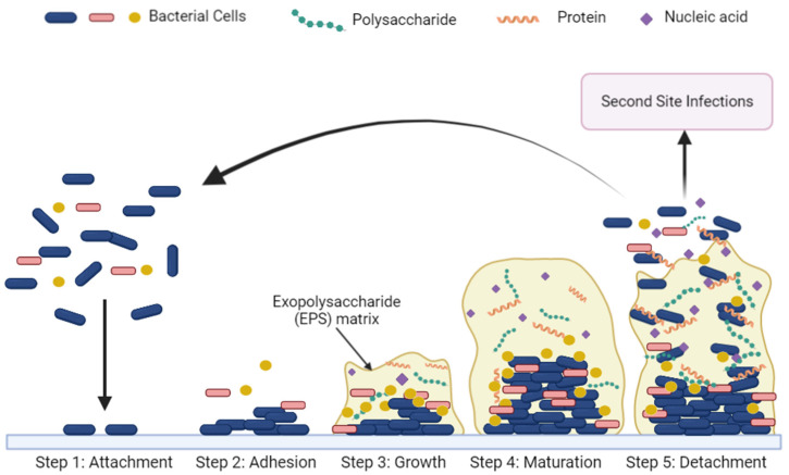
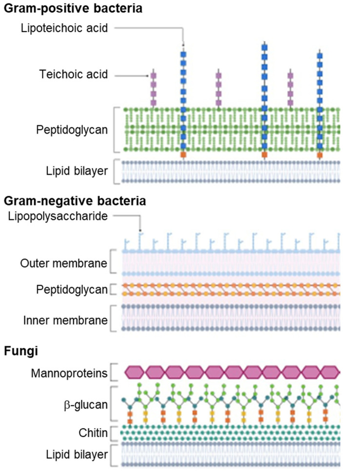
The rise of antibiotic resistant bacteria due to overuse and misuse of antibiotics in medicine and dentistry is a growing concern. New approaches are needed to combat antibiotic resistant (AR) bacterial infections. There are a number of methods available and in development to address AR infections. Dentists conventionally use chemicals such as chlorohexidine and calcium hydroxide to kill oral bacteria, with many groups recently developing more biocompatible antimicrobial peptides (AMPs) for use in the oral cavity. AMPs are promising candidates in the treatment of (oral) infections. Also known as host defense peptides, AMPs have been isolated from animals across all kingdoms of life and play an integral role in the innate immunity of both prokaryotic and eukaryotic organisms by responding to pathogens. Despite progress over the last four decades, there are only a few AMPs approved for clinical use. This review summarizes an Introduction to Oral Microbiome and Oral Infections, Traditional Antibiotics and Alternatives & Antimicrobial Peptides. There is a focus on cationic AMP characteristics and mechanisms of actions, and an overview of animal-derived natural and synthetic AMPs, as well as observed microbial resistance.

Keywords: AMPs; anti-microbial peptides; antibiotic resistance; microbiome; oral infections.


Conflict of interest statement

The authors declare no conflict of interest.

Figures

Figure 1
Figure 1
Progression of biofilm formation in the oral cavity. In the attachment stage (Step 1), planktonic cells begin to adhere to a surface. A monolayer of planktonic cells accumulates on the surface during the adhesion stage (Step 2). During the growth stage (Step 3), sessile cells begin to excrete an exopolysaccharide matrix containing proteins, polysaccharides (carbohydrates) and nucleic acids that surround the cells. The EPS matrix assumes a mushroom cloud shape in the maturation state (Step 4). Finally, planktonic cells are released from the EPS matrix during the detachment stage (Step 5), allowing for dispersal and colonization of new sites. (Created with BioRender, accessed on 1 August 2022).
Figure 2
Figure 2
Localization of bacterial oral pathogenesis. (1) Dental Caries: tooth enamel dissolved by acid produced by specific bacteria (e.g., Mutans streptococci, Lactobacilli). (2) Dental Pulp Infection: necrotic pulp tissues infected by microorganisms causing pulp inflammation and eventual necrosis. (3) Plaque and Calculus: biofilms form on non-shedding surfaces of the teeth; formation leads to caries and periodontal inflammation. (4) Periodontitis: dysbiotic bacterial infections cause inflammation that destroys periodontal ligament and bone. (5) Abscess: periodontal and periapical abscesses are pockets of pus caused by bacterial infection.
Figure 3
Figure 3
Cone beam computed tomography (CBCT) images detect the presence of apical inflammatory lesions. This type of empirical evidence for oral infections is used as a method of deciding treatment course by dentists. (A) CBCT sagittal image shows an apical inflammatory lesion as a darker grey pocket surrounding the apex of tooth. (B) CBCT images show dental caries from sagittal and cross-sectional views. (C) Sagittal CBCT image shows large apical inflammatory lesion between the apices of adjacent teeth. From the author’s collection.
Figure 4
Figure 4
Sources of AMPs from each of the six kingdoms, as of 18 October 2021, and secondary structures of representative AMPs. Data compiled from the Antimicrobial Peptide Database (https://aps.unmc.edu/, accessed on 1 August 2022). Basic residues are shown as orange, dark green represents aromatic residues, light green represents nonpolar residues and proline is shown in yellow. All structures were modeled using UniProt and predicted through either NMR, X-ray, or AlphaFold.
Figure 5
Figure 5
Overview of roles and observed mechanisms of action of antimicrobial peptides (created with BioRender.com, accessed on 1 August 2022). Either direct killing of bacteria and fungi, immunomodulation of host cells or a combination of both mechanisms are observed with AMPs.
Figure 6
Figure 6
Comparison of fungal and bacterial cells (created with BioRender.com, accessed on 1 August 2022). Gram-positive bacteria and fungi contain glycan layers on the extracellular side of the lipid membrane, while a second membrane encapsulates the peptidoglycan layer in Gram-negative bacteria.

Similar articles

Cited by

References

    1. Bik E.M., Long C.D., Armitage G.C., Loomer P., Emerson J., Mongodin E.F., Nelson K.E., Gill S.R., Fraser-Liggett C.M., Relman D.A. Bacterial diversity in the oral cavity of 10 healthy individuals. ISME J. 2010;4:962–974. doi: 10.1038/ismej.2010.30. - DOI - PMC - PubMed
    1. Ding T., Schloss P.D. Dynamics and associations of microbial community types across the human body. Nature. 2014;509:357–360. doi: 10.1038/nature13178. - DOI - PMC - PubMed
    1. Deo P.N., Deshmukh R. Oral microbiome: Unveiling the fundamentals. J. Oral Maxillofac. Pathol. 2019;23:122–128. doi: 10.4103/jomfp.JOMFP_304_18. - DOI - PMC - PubMed
    1. Burcham Z.M., Garneau N.L., Comstock S.S., Tucker R.M., Knight R., Metcalf J.L., Miranda A., Reinhart B., Meyers D., Woltkamp D., et al. Patterns of Oral Microbiota Diversity in Adults and Children: A Crowdsourced Population Study. Sci. Rep. 2020;10:2133. doi: 10.1038/s41598-020-59016-0. - DOI - PMC - PubMed
    1. Dominguez-Bello M.G., Costello E.K., Contreras M., Magris M., Hidalgo G., Fierer N., Knight R. Delivery mode shapes the acquisition and structure of the initial microbiota across multiple body habitats in newborns. Proc. Natl. Acad. Sci. USA. 2010;107:11971–11975. doi: 10.1073/pnas.1002601107. - DOI - PMC - PubMed