. 2022 Aug 15;14(8):1698.
doi: 10.3390/pharmaceutics14081698.

Efficient Delivery of DNA Using Lipid Nanoparticles

Affiliations

Efficient Delivery of DNA Using Lipid Nanoparticles

Lishan Cui et al. Pharmaceutics. .

Abstract

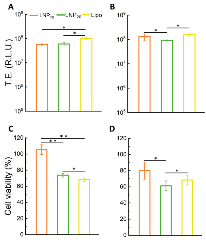
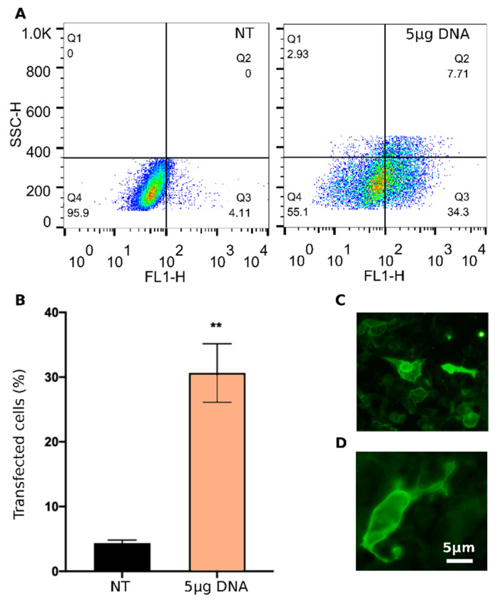
DNA vaccination has been extensively studied as a promising strategy for tumor treatment. Despite the efforts, the therapeutic efficacy of DNA vaccines has been limited by their intrinsic poor cellular internalization. Electroporation, which is based on the application of a controlled electric field to enhance DNA penetration into cells, has been the method of choice to produce acceptable levels of gene transfer in vivo. However, this method may cause cell damage or rupture, non-specific targeting, and even degradation of pDNA. Skin irritation, muscle contractions, pain, alterations in skin structure, and irreversible cell damage have been frequently reported. To overcome these limitations, in this work, we use a microfluidic platform to generate DNA-loaded lipid nanoparticles (LNPs) which are then characterized by a combination of dynamic light scattering (DLS), synchrotron small-angle X-ray scattering (SAXS), and transmission electron microscopy (TEM). Despite the clinical successes obtained by LNPs for mRNA and siRNA delivery, little is known about LNPs encapsulating bulkier DNA molecules, the clinical application of which remains challenging. For in vitro screening, LNPs were administered to human embryonic kidney 293 (HEK-293) and Chinese hamster ovary (CHO) cell lines and ranked for their transfection efficiency (TE) and cytotoxicity. The LNP formulation exhibiting the highest TE and the lowest cytotoxicity was then tested for the delivery of the DNA vaccine pVAX-hECTM targeting the human neoantigen HER2, an oncoprotein overexpressed in several cancer types. Using fluorescence-activated cell sorting (FACS), immunofluorescence assays and fluorescence confocal microscopy (FCS), we proved that pVAX-hECTM-loaded LNPs produce massive expression of the HER2 antigen on the cell membrane of HEK-293 cells. Our results provide new insights into the structure-activity relationship of DNA-loaded LNPs and pave the way for the access of this gene delivery technology to preclinical studies.

Keywords: DNA vaccines; HER2; lipid nanoparticles.


Conflict of interest statement

The authors declare no conflict of interest.

Figures

Figure 1
Figure 1
Schematic representation of the synthesis procedure of LNP DNA vaccines.
Figure 2
Figure 2
Size and zeta-potential of LNPs. (A) Size distributions, (B) mean size (reported as z-average) and polydispersity index (PdI) values, (C) zeta-potential distributions, and (D) average zeta-potential of LNP10 (orange) and LNP20 (green).
Figure 3
Figure 3
Nanostructure of LNPs. (A) Representative TEM image of LNP10 (panel A) (scale bar = 100 nm). (B) Synchrotron SAXS pattern of LNP10. The peaks arose from the lamellar periodicity of the system along the normal direction to the lipid bilayer. The small persistence length suggests that LNPs are made of randomly oriented lamellar domains, as schematically depicted in the inset. An average domain size made of 6 repeated units was estimated by relating the location of the first-ordered Bragg peak with its full width at half maximum (FWHM).
Figure 4
Figure 4
Transfection efficiency and cell viability of LNPs. Transfection efficiency (TE) of LNP10 and LNP20 expressed as relative light units (RLU) for HEK-293 (A) and CHO (B) cells. Cell viability of HEK-293 cells (C) and CHO cells (D) after treatment with LNP10 and LNP20 expressed as a percentage with respect to untreated cells. Statistical significance was evaluated using Student’s t-test: * p < 0.05; ** p < 0.01 (no asterisk means lack of significance). LipofectamineTM 3000 was used as a control.
Figure 5
Figure 5
Intracellular behavior of LNPs. (A) Average iMSD curves of LNP–Texas Red in CHO (red, n = 6) and HEK cells (grey, n = 6). y-Axis intercept derived by fitting (circles) represents the average dimension of LNP–Texas Red clusters adhering to the plasma membrane. (B) iMSD-derived diffusion coefficients of LNP–Texas Red in CHO (red) and HEK-293 (grey). Boxes represent 25th–75th percentiles; whiskers represent standard deviation. (C) iMSD-derived size of LNP–Texas Red clusters in CHO (red) and HEK-293 (grey). Boxes represent 25th–75th percentiles; whiskers represent maximum–minimum ranges; lines represent median values. (D) Exemplary images of CHO and HEK-293 cells labeled with Hoechst (blue, for nuclei) and incubated with LNP–Texas Red (red). Scale bar = 5 μm.
Figure 6
Figure 6
Transfection efficiency of LNP10 carrying the anti-HER2 DNA vaccine pVAX-hECTM. (A) Representative dot plots (increasing dot density from blue to red) and (B) bar graphs showing the percentage of HER2-positive HEK-293 cells at 48 h post-transfection with pVAX-hECTM encapsulated into LNP10 (5 μg DNA/well) in comparison with non-treated (NT) cells and analyzed by flow cytometry. Error bars represent average ± S.D. (n = 6) ** p < 0.001 unpaired t-test. (C,D) Fluorescence microscope photographs of HEK-293 cells at 48 h post-transfection with pVAX-hECTM delivered by LNP10 (10× and 40× magnifications in C and D, respectively). (NT: non-treated cells).

References

    1. Siegel R.L., Miller K.D., Fuchs H.E., Jemal A. Cancer statistics, 2022. CA Cancer J. Clin. 2022;72:7–33. doi: 10.3322/caac.21708. - DOI - PubMed
    1. Vanni G., Pellicciaro M., Materazzo M., Pedini D., Portarena I., Buonomo C., Perretta T., Rizza S., Pistolese C.A., Buonomo O.C. Advanced stages and increased need for adjuvant treatments in breast cancer patients: The effect of the one-year COVID-19 pandemic. Anticancer Res. 2021;41:2689–2696. doi: 10.21873/anticanres.15050. - DOI - PubMed
    1. Vardy J.L., Liew A., Warby A., Elder A., Keshet I., Devine R., Ouliaris C., Renton C., Tattersall M.H., Dhillon H.M. On the receiving end: Have patient perceptions of the side-effects of cancer chemotherapy changed since the twentieth century? Support. Care Cancer. 2022;30:3503–3512. doi: 10.1007/s00520-022-06804-1. - DOI - PMC - PubMed
    1. Kraehenbuehl L., Weng C.-H., Eghbali S., Wolchok J.D., Merghoub T. Enhancing immunotherapy in cancer by targeting emerging immunomodulatory pathways. Nat. Rev. Clin. Oncol. 2022;19:37–50. doi: 10.1038/s41571-021-00552-7. - DOI - PubMed
    1. Abbaspour M., Akbari V. Cancer vaccines as a targeted immunotherapy approach for breast cancer: An update of clinical evidence. Expert Rev. Vaccines. 2022;21:337–353. doi: 10.1080/14760584.2022.2021884. - DOI - PubMed