

Identification of Transferrin Receptor 1 (TfR1) Overexpressed in Lung Cancer Cells, and Internalization of Magnetic Au-CoFe2O4 Core-Shell Nanoparticles Functionalized with Its Ligand in a Cellular Model of Small Cell Lung Cancer (SCLC)
- PMID: 36015341
- PMCID: PMC9413248
- DOI: 10.3390/pharmaceutics14081715
Identification of Transferrin Receptor 1 (TfR1) Overexpressed in Lung Cancer Cells, and Internalization of Magnetic Au-CoFe2O4 Core-Shell Nanoparticles Functionalized with Its Ligand in a Cellular Model of Small Cell Lung Cancer (SCLC)
Abstract
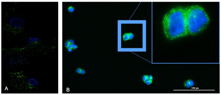
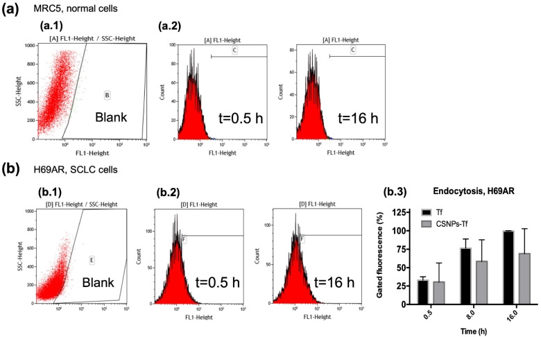
Lung cancer is, currently, one of the main malignancies causing deaths worldwide. To date, early prognostic and diagnostic markers for small cell lung cancer (SCLC) have not been systematically and clearly identified, so most patients receive standard treatment. In the present study, we combine quantitative proteomics studies and the use of magnetic core-shell nanoparticles (mCSNP's), first to identify a marker for lung cancer, and second to functionalize the nanoparticles and their possible application for early and timely diagnosis of this and other types of cancer. In the present study, we used label-free mass spectrometry in combination with an ion-mobility approach to identify 220 proteins with increased abundance in small cell lung cancer (SCLC) cell lines. Our attention was focused on cell receptors for their potential application as mCSNP's targets; in this work, we report the overexpression of Transferrin Receptor (TfR1) protein, also known as Cluster of Differentiation 71 (CD71) up to a 30-fold increase with respect to the control cell. The kinetics of endocytosis, evaluated by a flow cytometry methodology based on fluorescence quantification, demonstrated that receptors were properly activated with the transferrin supported on the magnetic core-shell nanoparticles. Our results are important in obtaining essential information for monitoring the disease and/or choosing better treatments, and this finding will pave the way for future synthesis of nanoparticles including chemotherapeutic drugs for lung cancer treatments.
Keywords: cluster of differentiation 71 (CD71); label-free; mass spectrometry; nanoparticles; small cell lung cancer (SCLC); transferrin receptor (TfR1).
Conflict of interest statement
The authors declare no conflict of interest. The funders had no role in the design of the study; in the collection, analyses, or interpretation of data; in the writing of the manuscript; or in the decision to publish the results.
Figures


Similar articles
-
Quantitative proteomic analysis of glycosylated proteins enriched from urine samples with magnetic ConA nanoparticles identifies potential biomarkers for small cell lung cancer.J Pharm Biomed Anal. 2021 Nov 30;206:114352. doi: 10.1016/j.jpba.2021.114352. Epub 2021 Aug 31. J Pharm Biomed Anal. 2021. PMID: 34509662
-
Aberrant expression of TfR1/CD71 in thyroid carcinomas identifies a novel potential diagnostic marker and therapeutic target.Thyroid. 2011 Mar;21(3):267-77. doi: 10.1089/thy.2010.0173. Epub 2011 Feb 16. Thyroid. 2011. PMID: 21323588
-
Depletion of ST6GALNACIII retards A549 non-small cell lung cancer cell proliferation by downregulating transferrin receptor protein 1 expression.Biochem Biophys Res Commun. 2021 Oct 20;575:78-84. doi: 10.1016/j.bbrc.2021.08.055. Epub 2021 Aug 25. Biochem Biophys Res Commun. 2021. PMID: 34461439
-
Antibody-mediated targeting of the transferrin receptor in cancer cells.Bol Med Hosp Infant Mex. 2016 Nov-Dec;73(6):372-379. doi: 10.1016/j.bmhimx.2016.11.004. Epub 2016 Dec 13. Bol Med Hosp Infant Mex. 2016. PMID: 29421281 Review.
-
Small cell lung cancer: etiology, biology, clinical features, staging, and treatment.Curr Probl Cancer. 1993 Mar-Apr;17(2):69-141. doi: 10.1016/0147-0272(93)90010-y. Curr Probl Cancer. 1993. PMID: 8395998 Review.
Cited by
-
Extracellular Vesicles for Clinical Diagnostics: From Bulk Measurements to Single-Vesicle Analysis.ACS Nano. 2025 Aug 12;19(31):28021-28109. doi: 10.1021/acsnano.5c00706. Epub 2025 Jul 28. ACS Nano. 2025. PMID: 40720603 Free PMC article. Review.
-
Opportunities and Challenges of Switchable Materials for Pharmaceutical Use.Pharmaceutics. 2022 Oct 28;14(11):2331. doi: 10.3390/pharmaceutics14112331. Pharmaceutics. 2022. PMID: 36365149 Free PMC article. Review.
-
A potential tumor marker: Chaperonin containing TCP‑1 controls the development of malignant tumors (Review).Int J Oncol. 2023 Sep;63(3):106. doi: 10.3892/ijo.2023.5554. Epub 2023 Aug 4. Int J Oncol. 2023. PMID: 37539774 Free PMC article. Review.
-
Elevated expression of transferrin receptor-1 in pancreatic cancer: clinical implications and prognostic significance.Clin Exp Med. 2025 Aug 28;25(1):307. doi: 10.1007/s10238-025-01847-0. Clin Exp Med. 2025. PMID: 40877641 Free PMC article.
-
Advances in Brain Tumor Therapy Based on the Magnetic Nanoparticles.Int J Nanomedicine. 2023 Dec 20;18:7803-7823. doi: 10.2147/IJN.S444319. eCollection 2023. Int J Nanomedicine. 2023. PMID: 38144513 Free PMC article. Review.
References
-
- Global Cancer Observatory: Cancer Today. World Health Organization; [(accessed on 12 June 2022)]. Available online: https://gco.iarc.fr/today.
-
- Wistuba I.I., Brambilla E., Noguchi M. IASLC Thoracic Oncology. Elsevier; Amsterdam, The Netherlands: 2018. Classic Anatomic Pathology and Lung Cancer.
LinkOut - more resources
Full Text Sources
