. 2022 Jul 27;14(8):1641.
doi: 10.3390/v14081641.

Ivermectin Inhibits the Replication of Usutu Virus In Vitro

Affiliations

Ivermectin Inhibits the Replication of Usutu Virus In Vitro

Maria Elisabeth Wald et al. Viruses. .

Abstract

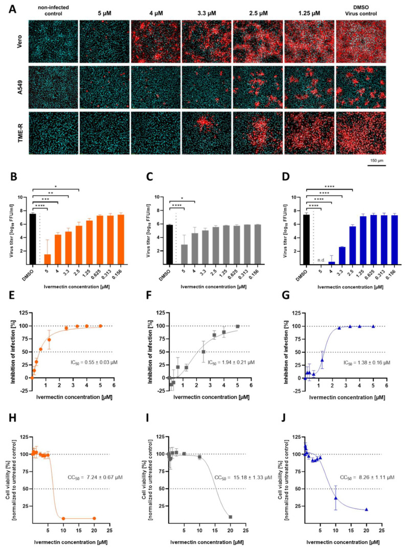
Usutu virus (USUV) is an emerging mosquito-borne arbovirus within the genus Flavivirus, family Flaviviridae. Similar to the closely related West Nile virus (WNV), USUV infections are capable of causing mass mortality in wild and captive birds, especially blackbirds. In the last few years, a massive spread of USUV was present in the avian population of Germany and other European countries. To date, no specific antiviral therapies are available. Nine different approved drugs were tested for their antiviral effects on the replication of USUV in vitro in a screening assay. Ivermectin was identified as a potent inhibitor of USUV replication in three cell types from different species, such as simian Vero CCL-81, human A549 and avian TME R. A 2- to 7-log10 reduction of the viral titer in the supernatant was detected at a non-cytotoxic concentration of 5 µM ivermectin dependent on the applied cell line. IC50 values of ivermectin against USUV lineage Africa 3 was found to be 0.55 µM in Vero CCL-81, 1.94 µM in A549 and 1.38 µM in TME-R cells. The antiviral efficacy was comparable between the USUV lineages Africa 2, Africa 3 and Europe 3. These findings show that ivermectin may be a candidate for further experimental and clinical studies addressing the treatment of USUV disease, especially in captive birds.

Keywords: Usutu virus; antiviral; bird; drug; ivermectin.


Conflict of interest statement

The authors declare no conflict of interest.

Figures

Figure 1
Figure 1
USUV replication kinetics varies in different cell lines. (A) Vero CCL-81, (B) A549 and (C) TME-R cell lines were infected with varying MOI doses (MOI of 0.01; MOI of 0.1; MOI of 1.0) of USUV lineage Africa 3 for 2 h. Parts from the supernatants were harvested every 24 h and viral infectivity was determined by focus-forming assay in Vero CCL-81. All experiments were performed in independent triplicates. Viral titer is depicted as mean log10 FFU/mL ± SD.
Figure 2
Figure 2
Screening of approved compounds for antiviral activity against USUV by titration of virus infectivity with a focus-forming assay. Several pharmacological molecules were screened for inhibitory efficacy against USUV lineage Africa 3 in reference cell line Vero CCL-81 at MOI of 0.1. (A) Extracellular infectious virus was quantified from supernatants by virus titration performing focus-forming assay. (B) Distribution of intracellular flavivirus antigen was evaluated by immunofluorescence analysis with a flavivirus-specific antibody. Flavivirus antigen is shown in red and nuclei stained with DAPI are shown in cyan blue. All results are demonstrated at the representative maximum concentration of 5 µM. Statistical significance is indicated as **** (p < 0.0001).
Figure 3
Figure 3
Antiviral efficacy of ivermectin against USUV in a concentration-dependent manner in several cell lines. Dose-dependent antiviral activity of ivermectin against USUV lineage Africa 3 at MOI of 0.1 was assessed in simian cell line Vero CCL-81, human cell line A549 and avian cell line TME-R. (A) Immunofluorescence staining of USUV-infected cells was performed in the presence of different ivermectin concentrations. Due to correlation between viral titer in the supernatant and percentage of infected cells, staining was done after 24 h in A549 and after 72 h in Vero CCL 81 and TME-R cells. Flavivirus antigen is depicted in red and cell nuclei stained with DAPI are shown in cyan blue. (BD) Quantification of extracellular infectious USUV particles released from Vero CCL-81 (B), A549 (C) and TME-R (D) was evaluated by virus titration. (EG) Half maximal inhibitory concentrations (IC50) of ivermectin were calculated in Vero CCL-81 (E), A549 (F) and TME-R (G) cells. Inhibition of infection was calculated by titration of supernatants from untreated USUV control in comparison to ivermectin treated panels. Data represent mean values ± SD from independent triplicates. (HJ) Viability of different cell lines in the presence of ivermectin. Cytotoxicity of ivermectin was measured in two-fold serial dilutions in cells lines Vero CCL 81 (H), A549 (I) and TME-R (J), applying a WST-8 tetrazolium salt system. Results are depicted as mean percentages ± SD of viable cells in comparison to the untreated control. Experiments were performed in independent triplicates. CC50 values were calculated by non-linear regression analysis performed in GraphPad Prism software 9. Statistical analysis was performed by ANOVA and Dunnett’s post-hoc multiple comparisons test evaluating IC50 values by non-linear regression analysis and indicating statistical significance as n.d. (not detectable, under detection limit), * (p < 0.05), ** (p < 0.01), *** (p < 0.001) and **** (p < 0.0001).
Figure 4
Figure 4
Antiviral activity of ivermectin against several USUV strains in vitro. TME-R cells were infected with USUV lineages Africa 3, Europe 3 and Africa 2 at MOI of 0.1 in the presence of selected ivermectin concentrations. Readout was performed after 72 h post infection. (A) Immunofluorescence staining of USUV-infected cells was performed in the presence of selected ivermectin concentrations. Flavivirus antigen is depicted in red and cell nuclei stained with DAPI are shown in cyan blue. (B) Virus titer was determined from supernatants of infected cells treated with 2.5 µM and 5 µM of ivermectin compared to vehicle control (DMSO). N.d. = not detectable. Data represent mean values ± SD from three independent experiments. Statistical analysis was performed by ANOVA and Dunnett’s post-hoc multiple comparisons test indicating statistical significance as n.d. (not detectable, under detection limit), ** (p < 0.01) and **** (p < 0.0001).

Similar articles

Cited by

References

    1. Vilibic-Cavlek T., Petrovic T., Savic V., Barbic L., Tabain I., Stevanovic V., Klobucar A., Mrzljak A., Ilic M., Bogdanic M., et al. Epidemiology of Usutu Virus: The European Scenario. Pathogens. 2020;9:699. doi: 10.3390/pathogens9090699. - DOI - PMC - PubMed
    1. Steinmetz H.W., Bakonyi T., Weissenböck H., Hatt J.-M., Eulenberger U., Robert N., Hoop R., Nowotny N. Emergence and establishment of Usutu virus infection in wild and captive avian species in and around Zurich, Switzerland-genomic and pathologic comparison to other central European outbreaks. Vet. Microbiol. 2011;148:207–212. doi: 10.1016/j.vetmic.2010.09.018. - DOI - PubMed
    1. Rijks J.M., Kik M.L., Slaterus R., Foppen R., Stroo A., IJzer J., Stahl J., Gröne A., Koopmans M., van der Jeugd H.P., et al. Widespread Usutu virus outbreak in birds in The Netherlands, 2016. Eurosurveillance. 2016;21:30391. doi: 10.2807/1560-7917.ES.2016.21.45.30391. - DOI - PMC - PubMed
    1. Sieg M., Schmidt V., Ziegler U., Keller M., Höper D., Heenemann K., Rückner A., Nieper H., Muluneh A., Groschup M.H., et al. Outbreak and Cocirculation of Three Different Usutu Virus Strains in Eastern Germany. Vector-Borne Zoonotic Dis. 2017;17:662–664. doi: 10.1089/vbz.2016.2096. - DOI - PubMed
    1. Cadar D., Lühken R., van der Jeugd H., Garigliany M., Ziegler U., Keller M., Lahoreau J., Lachmann L., Becker N., Kik M., et al. Widespread activity of multiple lineages of Usutu virus, western Europe, 2016. Eurosurveillance. 2017;22:30452. doi: 10.2807/1560-7917.ES.2017.22.4.30452. - DOI - PMC - PubMed

Publication types

Supplementary concepts

LinkOut - more resources