. 2022 Jul 27;14(8):1649.
doi: 10.3390/v14081649.

Characterization of a Human Sapovirus Genotype GII.3 Strain Generated by a Reverse Genetics System: VP2 Is a Minor Structural Protein of the Virion

Affiliations

Characterization of a Human Sapovirus Genotype GII.3 Strain Generated by a Reverse Genetics System: VP2 Is a Minor Structural Protein of the Virion

Tian-Cheng Li et al. Viruses. .

Abstract

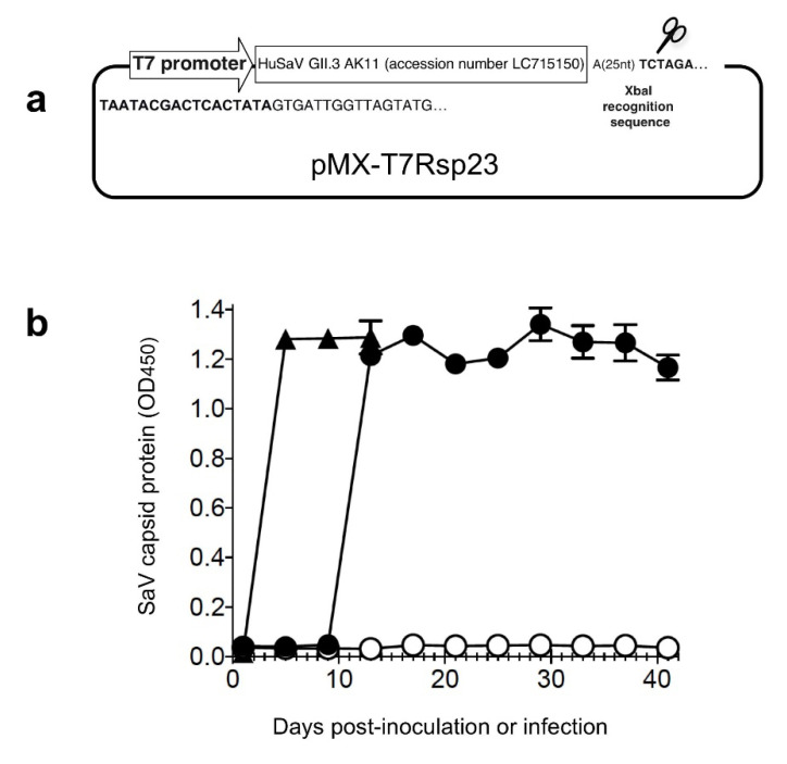
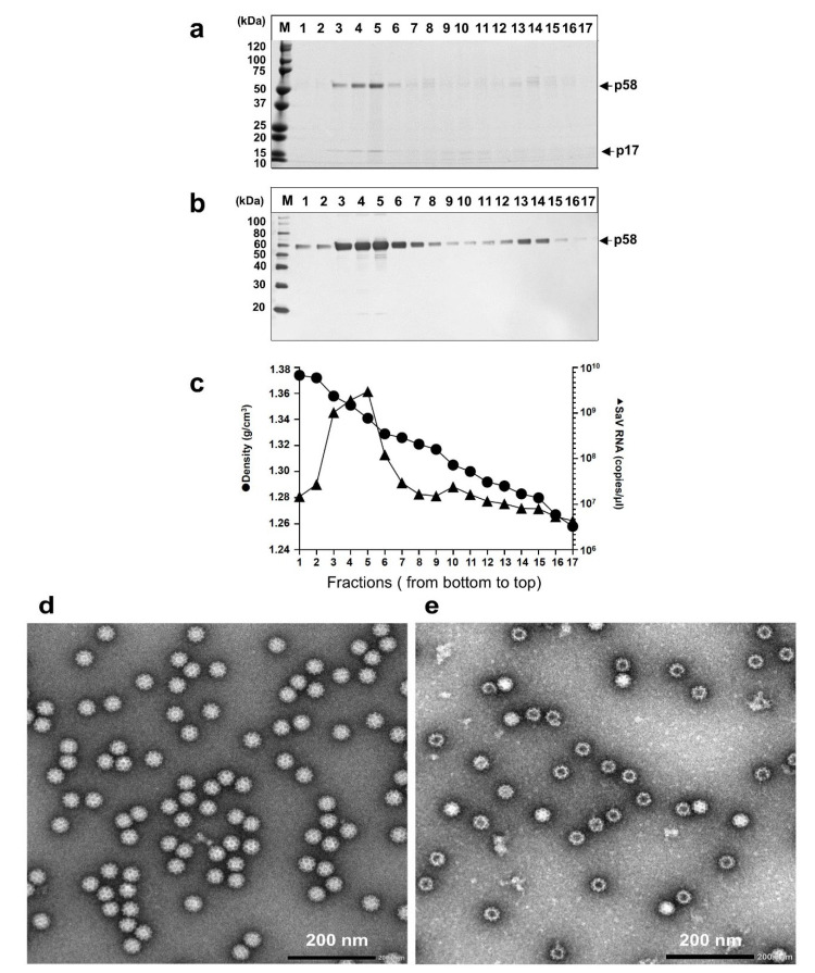
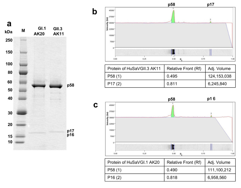
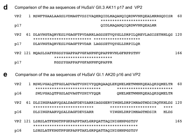
We devised a reverse genetics system to generate an infectious human sapovirus (HuSaV) GII.3 virus. Capped/uncapped full-length RNAs derived from HuSaV GII.3 AK11 strain generated by in vitro transcription were used to transfect HuTu80 human duodenum carcinoma cells; infectious viruses were recovered from the capped RNA-transfected cells and passaged in the cells. Genome-wide analyses indicated no nucleotide sequence change in the virus genomes in the cell-culture supernatants recovered from the transfection or those from the subsequent infection. No virus growth was detected in the uncapped RNA-transfected cells, suggesting that the 5'-cap structure is essential for the virus' generation and replication. Two types of virus particles were purified from the cell-culture supernatant. The complete particles were 39.2-nm-dia., at 1.350 g/cm3 density; the empty particles were 42.2-nm-dia. at 1.286 g/cm3. Two proteins (58-kDa p58 and 17-kDa p17) were detected from the purified particles; their molecular weight were similar to those of VP1 (~60-kDa) and VP2 (~16-kDa) of AK11 strain deduced from their amino acids (aa) sequences. Protein p58 interacted with HuSaV GII.3-VP1-specific antiserum, suggesting that p58 is HuSaV VP1. A total of 94 (57%) aa of p17 were identified by mass spectrometry; the sequences were identical to those of VP2, indicating that the p17 is the VP2 of AK11. Our new method produced infectious HuSaVs and demonstrated that VP2 is the minor protein of the virion, suggested to be involved in the HuSaV assembly.

Keywords: HuSaV; VP1; VP2; genogroup GII.3; human sapovirus; reverse genetics system; virus particle.


Conflict of interest statement

The authors declare no conflict of interest.

Figures

Figure 1
Figure 1
Generation and replication of HuSaV GII.3 AK11 strain in HuTu80 cells. A schematic diagram of the plasmid pMX-T7Rsp23 is shown (a). HuTu80 cells were transfected with either capped (⬤) or uncapped (⭘) full-length AK11 RNA, and the culture supernatant (p0) was collected every 4 days and used for detection of the capsid protein by an antigen-capture ELISA (b). The supernatant p0 collected on day 16 p.i. from the capped RNA-transfected cells was used to inoculate HuTu80 cells, and the capsid protein in the cell culture supernatant (p1) was examined similarly (▲) (b). Triplicate samples were used for transfection and inoculation. The mean OD value is shown and the error bars indicate the SD.
Figure 2
Figure 2
Purification and characterization of AK11 virions. The p1 supernatants collected on day 8 p.i. were concentrated by ultracentrifugation and then purified by CsCl equilibrium density gradient centrifugation. Aliquots from each fraction were analyzed by electrophoresis on 5–20% polyacrylamide gels and stained with CBB (a), and the capsid protein was detected by a Western blotting assay using a VP1-specific antiserum (b). Molecular weight markers (in kDa) are indicated on the left (a,b). Viral RNA in each fraction (▲) detected by RT-qPCR and the density (⬤) is shown (c). Electron micrographs of fractions 5 (d) and 13 (e). Bar: 200 nm. The particle sizes were determined using Hitachi EMIP software ver. 0524 (Hitachi High Technologies, Tokyo, Japan).
Figure 3
Figure 3
Proteins analyses of the p17 and p16. Purified HuSaV GI.1 AK20 virions and HuSaV GII.3 AK11 virions were analyzed by SDS-PAGE followed by CBB staining (a). The ratio of the protein content between p58 and p17 of HuSaV GII.3 AK11 (b), and that between p58 and p16 of HuSaV GI.1 AK20 (c) were quantitated by Image software ver. 6.1 based on the band intensities. The identified aa sequences of p17 was compared with those of VP2 of HuSaV GII.3 AK11 (d), and the aa sequences of p16 was compared with those of VP2 of HuSaV GI.1AK20 (e).
Figure 3
Figure 3
Proteins analyses of the p17 and p16. Purified HuSaV GI.1 AK20 virions and HuSaV GII.3 AK11 virions were analyzed by SDS-PAGE followed by CBB staining (a). The ratio of the protein content between p58 and p17 of HuSaV GII.3 AK11 (b), and that between p58 and p16 of HuSaV GI.1 AK20 (c) were quantitated by Image software ver. 6.1 based on the band intensities. The identified aa sequences of p17 was compared with those of VP2 of HuSaV GII.3 AK11 (d), and the aa sequences of p16 was compared with those of VP2 of HuSaV GI.1AK20 (e).

References

    1. Oka T., Wang Q., Katayama K., Saif L.J. Comprehensive review of human sapoviruses. Clin. Microbiol. Rev. 2015;28:32–53. doi: 10.1128/CMR.00011-14. - DOI - PMC - PubMed
    1. Oka T., Lu Z., Phan T., Delwart E.L., Saif L.J., Wang Q. Genetic Characterization and Classification of Human and Animal Sapoviruses. PLoS ONE. 2016;11:e0156373. doi: 10.1371/journal.pone.0156373. - DOI - PMC - PubMed
    1. Yinda C.K., Conceição-Neto N., Zeller M., Heylen E., Maes P., Ghogomu S.M., Van Ranst M., Matthijnssens J. Novel highly divergent sapoviruses detected by metagenomics analysis in straw-colored fruit bats in Cameroon. Emerg. Microbes Infect. 2017;6:e38. doi: 10.1038/emi.2017.20. - DOI - PMC - PubMed
    1. Li J., Zhang W., Cui L., Shen Q., Hua X. Metagenomic identification, genetic characterization and genotyping of porcine sapoviruses. Infect. Genet. Evol. 2018;62:244–252. doi: 10.1016/j.meegid.2018.04.034. - DOI - PubMed
    1. Hansman G.S., Oka T., Sakon N., Takeda N. Antigenic diversity of human sapoviruses. Emerg. Infect. Dis. 2007;13:1519–1525. doi: 10.3201/eid1310.070402. - DOI - PMC - PubMed

Publication types

LinkOut - more resources