. 2022 Aug 1;14(8):1701.
doi: 10.3390/v14081701.

Evolutionary Conservation of PP2A Antagonism and G2/M Cell Cycle Arrest in Maedi-Visna Virus Vif

Affiliations

Evolutionary Conservation of PP2A Antagonism and G2/M Cell Cycle Arrest in Maedi-Visna Virus Vif

Adeline M Luperchio et al. Viruses. .

Abstract

The canonical function of lentiviral Vif proteins is to counteract the mutagenic potential of APOBEC3 antiviral restriction factors. However, recent studies have discovered that Vif proteins from diverse HIV-1 and simian immunodeficiency virus (SIV) isolates degrade cellular B56 phosphoregulators to remodel the host phosphoproteome and induce G2/M cell cycle arrest. Here, we evaluate the conservation of this activity among non-primate lentiviral Vif proteins using fluorescence-based degradation assays and demonstrate that maedi-visna virus (MVV) Vif efficiently degrades all five B56 family members. Testing an extensive panel of single amino acid substitution mutants revealed that MVV Vif recognizes B56 proteins through a conserved network of electrostatic interactions. Furthermore, experiments using genetic and pharmacologic approaches demonstrate that degradation of B56 proteins requires the cellular cofactor cyclophilin A. Lastly, MVV Vif-mediated depletion of B56 proteins induces a potent G2/M cell cycle arrest phenotype. Therefore, remodeling of the cellular phosphoproteome and induction of G2/M cell cycle arrest are ancient and conserved functions of lentiviral Vif proteins, which suggests that they are advantageous for lentiviral pathogenesis.

Keywords: HIV-1; MVV; PPP2R5; Vif; host-pathogen; phosphatase regulation.


Conflict of interest statement

The authors declare no conflict of interest.

Figures

Figure 1
Figure 1
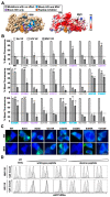
MVV Vif efficiently targets the family of B56α-ε proteins for degradation. (A,B) low cytometry histograms of A3-eGFP or eGFP-B56α degradation in the presence of the indicated Vif protein. The open histogram (black) represents the eGFP profile of cells expressing mTagBFP2 alone, whereas the filled histogram (grey) represents the profile of cells expressing the indicated Vif construct. The data shown are from one of three independent experiments. (C) Quantification of eGFP mean fluorescence intensity from cells expressing the indicated Vif proteins. ns, indicates no significance; * is p < 0.05 by unpaired student’s t-test. All experiments were repeated at least three independent times.
Figure 2
Figure 2
CPYA is required for MVV Vif-induce degradation of B56 proteins. (A,B) Flow cytometry histograms were generated as described in Figure 1. For cyclosporin A (CsA) treatments, the indicated concentration of inhibitor was added to samples transiently expressing either HIV or MVV Vif proteins. (C) Quantification of eGFP mean fluorescence intensity from cells expressing the indicated Vif proteins with or without increasing concentration of CsA treatment. *** is p < 0.001 by an unpaired student’s t-test. (D) RT-PCR analysis of HEK293FT eGFP-B56α cells stably expressing the indicated shRNA control or CYPA knock-down constructs. (E) Flow cytometry histograms were generated as described in Figure 1. Dashed lines represent eGFP-B56α cells expressing a shRNA control vector and solid lines represent eGFP-B56α cells expressing a CYPA knock-down construct. All experiments were repeated at least three independent times.
Figure 3
Figure 3
HIV and MVV Vif proteins partially distinct negatively charged B56 surfaces. (A) Left, surface depiction of B56γ (PDB: 2IAE) with residues required or dispensable for Vif degradation highlighted in the indicated color. Grey, no impact to degradation; purple, inhibit HIV Vif-induced degradation; blue, inhibit both HIV and MVV Vif-induced degradation; orange, peptide inhibitor binding site. Right, electrostatic surface potential map with red indicating negative charge, white indicating neutral charge, and blue indicating positive charge. Vif binding surfaces depicted by dashed lines (B) Quantification of eGFP mean fluorescence intensity from eGFP-B56α cells expressing the indicated Vif proteins. No statistical value indicates no significant difference between respective wild-type and mutant Vif proteins; * is p < 0.05; ** is p < 0.01; *** is p < 0.001 by an unpaired student’s t-test. (C) Fluorescence microscopy images of HEK293T cells transiently expressing the indicated Vif proteins and B56α proteins; scale bar is 10 μM (D) Flow cytometry histograms were generated as described in Figure 1. Cells were co-transfected with the indicated Vif protein with either wild-type (LxxIxE) or alanine mutant (AxxAxA) B56 inhibitor peptide constructs. All experiments were repeated at least three independent times.
Figure 4
Figure 4
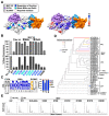
B56 antagonism and G/2M cell cycle arrest are conserved activities in global MVV Vif isolates. (A) Left, computational model of MVV Vif in complex with CPYA (PDB:1CWA) and ELOB/C (PDB:4N9F). Surface arginine or lysine residues that are required for MVV Vif-induced degradation of eGFP-B56α are colored based on separation-of-function (blue) or complete loss of degradation activity (cyan). Right, electrostatic surface potential map with red indicating negative charge, white indicating neutral charge, and blue indicating positive charge. Arginine and lysine residues are highlighted by dashed red outlines. (B) Quantification of eGFP mean fluorescence intensity from eGFP-B56α cells expressing the indicated MVV Vif proteins. (C) Weblogo of MVV Vif sequences downloaded from the NCBI database with amino acid residues required for the indicated protein–protein interactions highlighted (n = 40). Frequency of amino acid polymorphisms at all positions that exhibited variability are depicted as a bar graph (right). (D) Phylogenetic analysis of MVV Vif sequences with corresponding accession number and geographic region of isolation depicted. Coloring indicates the number of amino acid polymorphisms at positions required for MVV Vif-induced degradation of B56α-eGFP. Red, 4 amino acid polymorphisms; orange, 2 amino acid polymorphisms; purple, 1 amino acid polymorphism; grey, no polymorphisms. The blue asterisk highlights the MVV Vif isolate used in this study. (E) Flow cytometry histograms of representative cell cycle profiles of HeLa cells transiently expressing the indicated wild-type or mutant Vif proteins. All experiments were repeated at least three independent times.

Similar articles

Cited by

References

    1. Harris R., Dudley J. APOBECs and virus restriction. Virology. 2015;479–480:131–145. doi: 10.1016/j.virol.2015.03.012. - DOI - PMC - PubMed
    1. Desimmie B.A., Delviks-Frankenberrry K.A., Burdick R.C., Qi D., Izumi T., Pathak V.K. Multiple APOBEC3 restriction factors for HIV-1 and one Vif to rule them all. J. Mol. Biol. 2014;426:1220–1245. doi: 10.1016/j.jmb.2013.10.033. - DOI - PMC - PubMed
    1. Malim M.H., Emerman M. HIV-1 accessory proteins--ensuring viral survival in a hostile environment. Cell Host Microbe. 2008;3:388–398. doi: 10.1016/j.chom.2008.04.008. - DOI - PubMed
    1. Simon V.B.N., Landau N. Intrinsic host restrictions to HIV-1 and mechanisms of viral escape. Nat. Immunol. 2015;16:546–553. doi: 10.1038/ni.3156. - DOI - PMC - PubMed
    1. Guo Y., Dong L., Qiu X., Wang Y., Zhang B., Liu H., Yu Y., Zang Y., Yang M., Huang Z. Structural basis for hijacking CBF-β and CUL5 E3 ligase complex by HIV-1 Vif. Nature. 2014;505:229–233. doi: 10.1038/nature12884. - DOI - PubMed

Publication types

Substances

LinkOut - more resources