Review
. 2022 Aug 9;14(8):1744.
doi: 10.3390/v14081744.

PEDV: Insights and Advances into Types, Function, Structure, and Receptor Recognition

Affiliations
Review

PEDV: Insights and Advances into Types, Function, Structure, and Receptor Recognition

Feng Lin et al. Viruses. .

Abstract

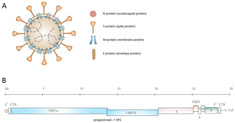
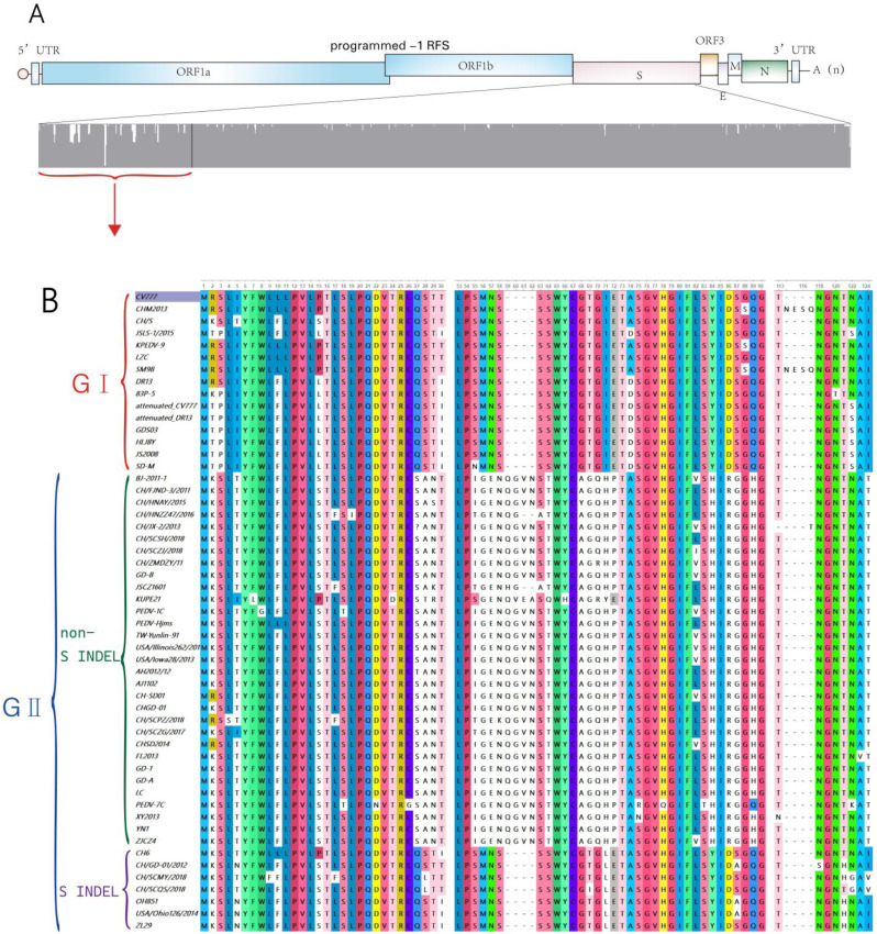
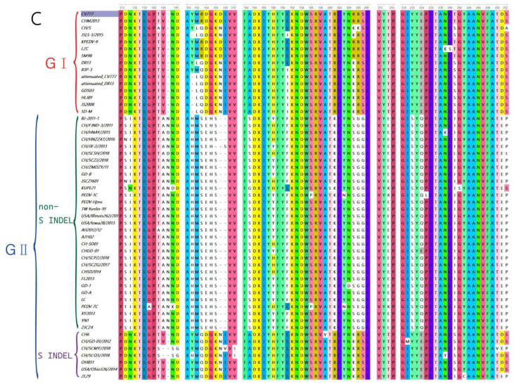
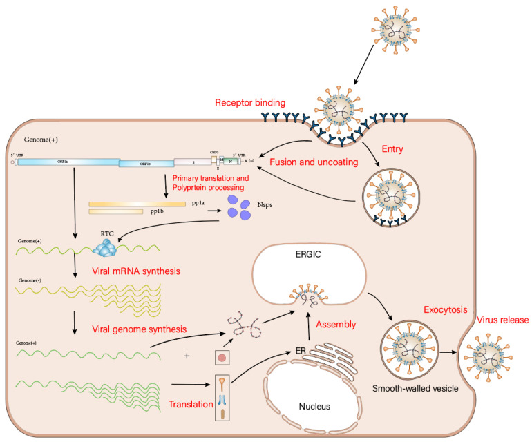
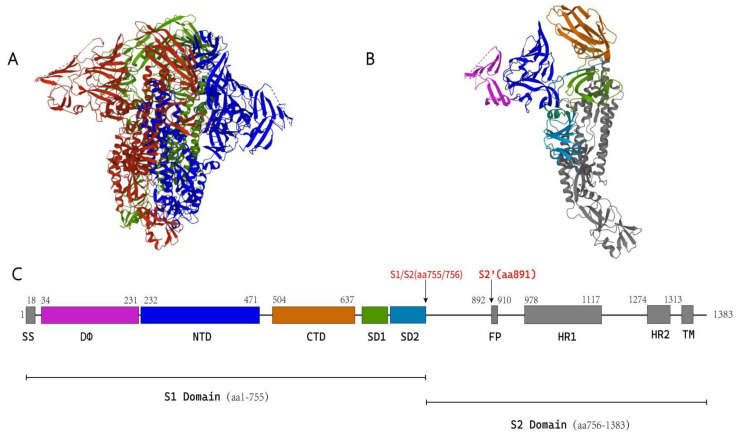
Porcine epidemic diarrhea virus (PEDV) has been endemic in most parts of the world since its emergence in the 1970s. It infects the small intestine and intestinal villous cells, spreads rapidly, and causes infectious intestinal disease characterized by vomiting, diarrhea, and dehydration, leading to high mortality in newborn piglets and causing massive economic losses to the pig industry. The entry of PEDV into cells is mediated by the binding of its spike protein (S protein) to a host cell receptor. Here, we review the structure of PEDV, its strains, and the structure and function of the S protein shared by coronaviruses, and summarize the progress of research on possible host cell receptors since the discovery of PEDV.

Keywords: PEDV receptor; PEDV variant strain; porcine epidemic diarrhea virus (PEDV); spike protein (S protein).


Conflict of interest statement

The authors declare that they have no conflict of interest.

Figures

Figure 1
Figure 1
Schematic representation of PEDV genome organization and virion structure. (A) Schematic diagram showing the structure of the virus. The lipid bilayer containing the S, M, and E proteins covers the core structure of the viral RNA genome, which is bound to the N protein, forming a long helical ribonucleoprotein (RNP) complex. (B) Structure of the RNA genome of PEDV. The RNA genome of PEDV is 28 kb in size and has a 5′ end cap and a 3′ polyadenylated tail. The viral genome is flanked by UTRs and contains seven open reading frames: ORF1a, ORF1b, S, ORF3, E, M, and N; these are indicated by the boxes. (PEDV’s ORF1a and ORF1b have an overlapping region within which the programmed –1 ribosomal frameshift (RFS) occurs).
Figure 2
Figure 2
(A) The amino acid sequence differences of S proteins of different PEDV strains are shown, with the differences mainly concentrated in the N-terminal of S protein. (B,C) Amino acid sequences of the highly variable N-terminal region of the S proteins of various PEDV strains are shown in detail; the different genetic subgroups are indicated by brackets of different colors. The G2 non-S INDEL PEDV strain has a different genetic profile than CV777, while the G2 S INDEL PEDV strain has a higher similarity to CV777.
Figure 2
Figure 2
(A) The amino acid sequence differences of S proteins of different PEDV strains are shown, with the differences mainly concentrated in the N-terminal of S protein. (B,C) Amino acid sequences of the highly variable N-terminal region of the S proteins of various PEDV strains are shown in detail; the different genetic subgroups are indicated by brackets of different colors. The G2 non-S INDEL PEDV strain has a different genetic profile than CV777, while the G2 S INDEL PEDV strain has a higher similarity to CV777.
Figure 3
Figure 3
Overview of the PEDV life cycle.
Figure 4
Figure 4
(A) The trimeric pre-fusion structure of the PEDV S protein is shown, with different colors rep-resenting different monomers. (B) Showing the structure of a monomer of the PEDV S protein. (C) Schematic diagram of the structure of a monomer of the PEDV S protein. Different colored boxes are used to represent the different structural domains of PEDV S proteins; the same color in di-agrams (B,C) indicates the same structural domain.

Similar articles

Cited by

References

    1. Pensaert M.B., de Bouck P. A new coronavirus-like particle associated with diarrhea in swine. Arch. Virol. 1978;58:243–247. doi: 10.1007/BF01317606. - DOI - PMC - PubMed
    1. Cavanagh D. Nidovirales: A new order comprising Coronaviridae and Arteriviridae. Arch. Virol. 1997;142:629–633. - PubMed
    1. Wood E. An apparently new syndrome of porcine epidemic diarrhoea. Veter. Rec. 1977;100:243–244. doi: 10.1136/vr.100.12.243. - DOI - PubMed
    1. Kocherhans R., Bridgen A., Ackermann M., Tobler K. Completion of the porcine epidemic diarrhoea coronavirus (PEDV) genome sequence. Virus Genes. 2001;23:137–144. doi: 10.1023/A:1011831902219. - DOI - PMC - PubMed
    1. Duarte M., Tobler K., Bridgen A., Rasschaert D., Ackermann M., Laude H. Sequence analysis of the porcine epidemic diarrhea virus genome between the nucleocapsid and spike protein genes reveals a polymorphic ORF. Virology. 1994;198:466–476. doi: 10.1006/viro.1994.1058. - DOI - PMC - PubMed

MeSH terms

Substances

LinkOut - more resources