. 2022 Aug 15;14(8):1782.
doi: 10.3390/v14081782.

Porcine Enteric Coronavirus PEDV Induces the ROS-ATM and Caspase7-CAD-γH2AX Signaling Pathways to Foster Its Replication

Affiliations

Porcine Enteric Coronavirus PEDV Induces the ROS-ATM and Caspase7-CAD-γH2AX Signaling Pathways to Foster Its Replication

Xin Ming et al. Viruses. .

Abstract

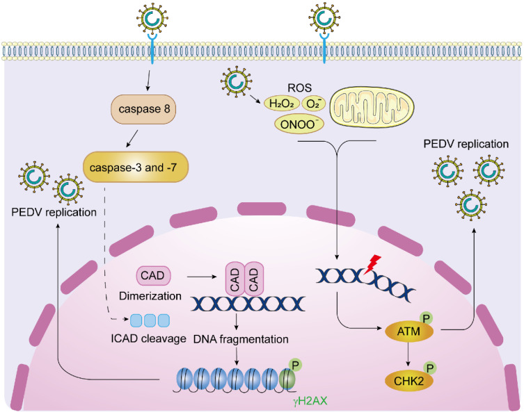
DNA damage response (DDR) is an evolutionarily conserved mechanism by which eukaryotic cells sense DNA lesions caused by intrinsic and extrinsic stimuli, including virus infection. Although interactions between DNA viruses and DDR have been extensively studied, how RNA viruses, especially coronaviruses, regulate DDR remains unknown. A previous study showed that the porcine epidemic diarrhea virus (PEDV), a member of the genus Alphacoronavirus in the Coronaviridae family, induces DDR in infected cells. However, the underlying mechanism was unclear. This study showed that PEDV activates the ATM-Chk2 signaling, while inhibition of ATM or Chk2 dampens the early stage of PEDV infection. Additionally, we found that PEDV-activated ATM signaling correlates with intracellular ROS production. Interestingly, we showed that, unlike the typical γH2AX foci, PEDV infection leads to a unique γH2AX staining pattern, including phase I (nuclear ring staining), II (pan-nuclear staining), and III (co-staining with apoptotic bodies), which highly resembles the apoptosis process. Furthermore, we demonstrated that PEDV-induced H2AX phosphorylation depends on the activation of caspase-7 and caspase-activated DNAse (CAD), but not ATM-Chk2. Finally, we showed that the knockdown of H2AX attenuates PEDV replication. Taken together, we conclude that PEDV induces DDR through the ROS-ATM and caspase7-CAD-γH2AX signaling pathways to foster its early replication.

Keywords: ATM; DNA damage response; PEDV; caspase; γH2AX.


Conflict of interest statement

The authors declare no conflict of interest.

Figures

Figure 1
Figure 1
PEDV infection activates ATM-dependent DNA damage response. (A) Western blots were prepared with extracts from Vero-E6 cells uninfected or infected with CV777 (0.1 MOI) for 12, 24, 36, and 48 h. The levels of p-ATM (S1981), total ATM, p-Chk2(T68), total Chk2, p-ATR(T1989), total ATR, p-DNA-PK(S2056), total DNA-PK, γH2AX, PEDV N, and actin were determined using their respective antibodies. Vero-E6 cells infected with UV-inactivated CV777 (0.1 MOI) for 48 h were used as the negative control. (B) Western blots were prepared with extracts from Vero-E6 cells uninfected or infected with HLJBY (0.1 MOI) for 12, 24, and 36 h. (C) Western blots were prepared with extracts from Marc145 cells uninfected or infected with CV777 (0.1 MOI) for 24, 36, and 48 h at 0.1 MOI.
Figure 2
Figure 2
Suppression of the ATM signaling pathway inhibits the replication of PEDV. (A,B) Western blots were prepared with extracts from Vero-E6 cells pretreated with DMSO, ATM antagonist KU55933 (6 μM) (A), or ATR antagonist VE821 (2 μM) (B) for 2 h and followed by infection with CV777 (0.5 MOI) in the presence of DMSO or KU55933 (A), or VE821 (B) for 12, 24, 30 h. (C) The virus titers were measured by PFU assay with supernatants harvested from Vero-E6 cells pretreated with DMSO or KU55933 (6 μM) or VE821 (2 μM) for 2 h before and during infection with CV777 (0.1 MOI) for 12 and 24 h. (D,E) Western blots were prepared with extracts from Vero-E6 cells transfected with scramble or 50 nM siATM (D), or siChk2 (E) for 48 h and followed by infection with 0.1 MOI CV777 for 6 and 12 h. (F,G) The experiment was performed as in (D,E), except the cells and supernatants were collected for PFU assay (* p < 0.05; ** p < 0.01; *** p < 0.001).
Figure 3
Figure 3
PEDV-induced cellular ROS contributes to the activation of the ATM signaling. (A) ROS levels were measured with Vero-E6 cells uninfected or infected with CV777 (1 MOI) for 12, 24, and 30 h. The relative ROS levels were calculated by the DCF fluorescence intensity in PEDV-infected cells. Results are representative of three independent experiments. (B) ROS levels were measured with Vero-E6 cells uninfected or infected with HLJBY (1 MOI) at 18, 24, and 30 h. (C) The experiment was performed as in A, except using Marc145 cells. (D) ROS levels were measured in Vero-E6 cells pre-treated with DMSO, APO (1 mM), NAC (100 μM), or DPI (0.5 μM) for 2 h and followed by CV777 (1 MOI) infection in the presence of DMSO, APO (1 mM), NAC (100 μM), or DPI (0.5 μM) for 24 h. (E) The quantification of the relative ROS levels calculated by the DCF fluorescence intensity in cells that were treated as in (D). Results are representative of three independent experiments. (F,G) Western blots were prepared with extracts from Vero-E6 cells treated with DMSO, APO (1 mM), NAC (100 μM), or DPI (0.5 μM) for 2 h before and during infection with CV777 (1 MOI) for 24 and 30 h (*** p < 0.001).
Figure 4
Figure 4
The pan-nuclear staining of γH2AX in PEDV-infected Vero-E6 cells. (A) Confocal images are representative of γH2AX immunofluorescence staining in Vero-E6 cells infected with PEDV (1 MOI) for 30 h. γH2AX was labeled in green, PEDV N was labeled in red, and nuclei were stained in blue with DAPI. (B) Representative confocal images of a single cell showing the γH2AX patterns in different phases (600× magnification). From left to right: untreated cells, etoposide-treated cells, and CV777 infected cells. The graph shows the relative distribution of the different γH2AX patterns (over 100 cells). Black columns correspond to peripheral nuclear staining (ring pattern, I), dark gray columns correspond to pan-staining (flooded pattern, II), and light gray columns correspond to apoptotic bodies fully stained with γH2AX (III).
Figure 5
Figure 5
PEDV-induced phosphorylation of H2AX is decreased by caspase inhibitors. (A) Western blots were prepared with extracts from Vero-E6 cells treated with DMSO or caspase pan-inhibitor Z-VAD-FMK (0, 10, and 20 μM) for 2 h before and during infection with CV777 (0.5 MOI) for 30 h. (B) Western blots were prepared with extracts from Vero-E6 cells treated with DMSO or caspase-8 inhibitor Z-IETD-FMK (50 μM), or caspase-3/7 inhibitor Ac-DEVD-CHO (50 μM) for 2 h before and during infection with CV777 (0.5 MOI) for 30 h. (C) IFA was performed with Vero-E6 cells treated with DMSO, Z-VAD-FMK (10 μM), Z-IETD-FMK (50 μM), and Ac-DEVD-CHO (50 μM) for 2 h, respectively, before and during infection with CV777 (0.5 MOI) for 30 h. The cells were fixed and double-immunostained with specific rabbit anti-γH2AX and mouse anti-N antibodies. The nuclei were stained with DAPI. The graph shows the percentage of γH2AX positive cells in about 200 PEDV-infected cells. (D,E) Western blots were prepared with extracts from Vero-E6 cells transiently transfected with 50 nM scramble or caspase-3/7 siRNA for 48 h, followed by 1 MOI CV777 infection for 25 h (** p < 0.01; *** p < 0.001).
Figure 6
Figure 6
CAD implicates in PEDV-induced H2AX phosphorylation. (A,B) Western blots were prepared with extracts from Vero-E6 cells transiently transfected with empty vector or Flag-tagged ICAD or ICAD-M (D117E, D224E) for 30 h, followed by 1 MOI CV777 (A) or HLJBY (B) infection for 25 h. (C) IFA was performed with Vero-E6 cells transiently transfected with empty vector or Flag-tagged ICAD or ICAD-M (D117E, D224E) for 30 h, followed by 1 MOI CV777 infection for 25 h. (D,E) Western blots were prepared with extracts from Vero-E6 cells transiently transfected with 50 nM scramble or CAD siRNA for 48 h, followed by 1 MOI CV777 (D) or HLJBY (E) infection for 25 and 30 h.
Figure 7
Figure 7
Silencing H2AX decreases PEDV replication. (A,B) Western blots were prepared with extracts from Vero-E6 cells transiently transfected with 50 nM scramble or H2AX siRNA for 48 h, followed by 1 MOI CV777 (A) or HLJBY (B) infection for 6 and 12 h. (C) Western blots were prepared with extracts from Vero-E6 cells transfected with 50 nM scramble or H2AX siRNA for 48 h, followed by 1 MOI CV777 infection for 6 and 12 h. The cells and supernatants were collected for PFU assay (** p < 0.01; *** p < 0.001).
Figure 8
Figure 8
The interplay between PEDV replication and cellular DNA damage response. After PEDV enters host cells and replicates in the cytoplasm, the accumulation of massive intracellular ROS activates ATM-mediated DSB. Activated ATM and its downstream substrate Chk2 facilitate viral replication in the early infection stage. In the late stage of PEDV infection, the initiator caspase-8 activates the executioner caspase-3 and caspase-7 which subsequently cleave ICAD and release CAD from the ICAD-CAD complex and promote the homodimerization of CAD. The scissor-like CAD dimers create DSBs in the genome and DNA fragmentation, leading to H2AX phosphorylation in the nucleus. Inhibition of H2AX, ATM, and Chk2 dampens the replication of PEDV.

Similar articles

Cited by

References

    1. Zhou B.B., Elledge S.J. The DNA damage response: Putting checkpoints in perspective. Nature. 2000;408:433–439. doi: 10.1038/35044005. - DOI - PubMed
    1. Roos W.P., Kaina B. DNA damage-induced cell death by apoptosis. Trends Mol. Med. 2006;12:440–450. doi: 10.1016/j.molmed.2006.07.007. - DOI - PubMed
    1. Sulli G., Di Micco R., di Fagagna F.D.A. Crosstalk between chromatin state and DNA damage response in cellular senescence and cancer. Nat. Rev. Cancer. 2012;12:709–720. doi: 10.1038/nrc3344. - DOI - PubMed
    1. Harper J.W., Elledge S.J. The DNA damage response: Ten years after. Mol. Cell. 2007;28:739–745. doi: 10.1016/j.molcel.2007.11.015. - DOI - PubMed
    1. Ryan E.L., Hollingworth R., Grand R.J. Activation of the DNA Damage Response by RNA Viruses. Biomolecules. 2016;6:2. doi: 10.3390/biom6010002. - DOI - PMC - PubMed

Publication types

Substances