. 2022 Jul 26;1(3):pgac142.
doi: 10.1093/pnasnexus/pgac142. eCollection 2022 Jul.

Frataxin controls ketone body metabolism through regulation of OXCT1

Affiliations

Frataxin controls ketone body metabolism through regulation of OXCT1

Yi Na Dong et al. PNAS Nexus. .

Abstract

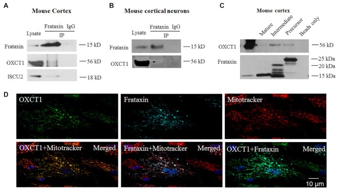
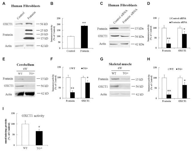
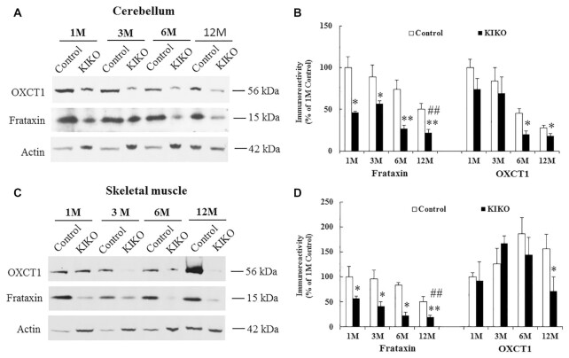
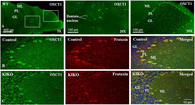
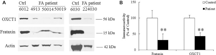
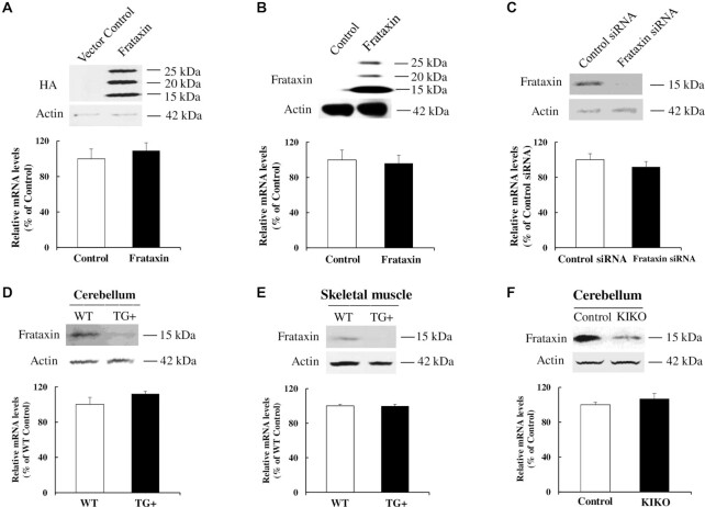
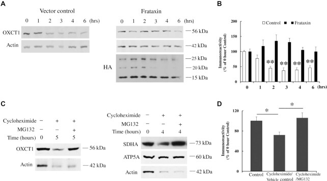
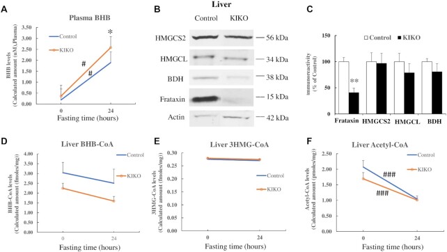
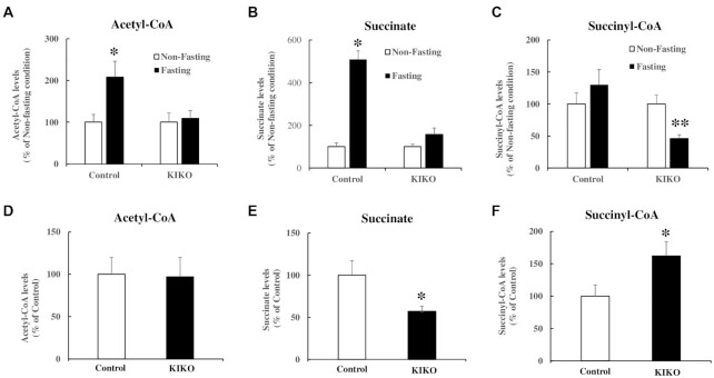
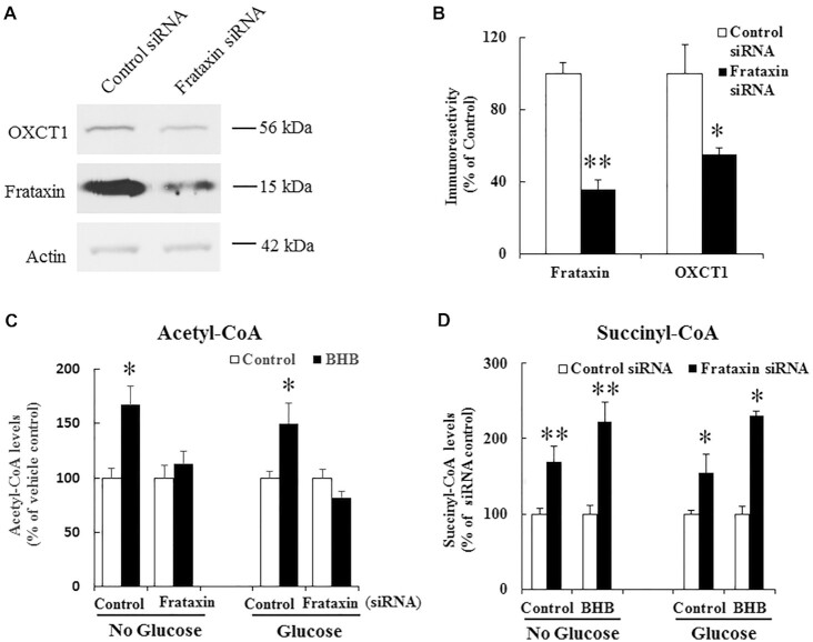
Friedreich's ataxia (FRDA) is an autosomal recessive neurodegenerative disease caused by the deficiency of mitochondrial protein frataxin, which plays a crucial role in iron-sulphur cluster formation and ATP production. The cellular function of frataxin is not entirely known. Here, we demonstrate that frataxin controls ketone body metabolism through regulation of 3-Oxoacid CoA-Transferase 1 (OXCT1), a rate limiting enzyme catalyzing the conversion of ketone bodies to acetoacetyl-CoA that is then fed into the Krebs cycle. Biochemical studies show a physical interaction between frataxin and OXCT1 both in vivo and in vitro. Frataxin overexpression also increases OXCT1 protein levels in human skin fibroblasts while frataxin deficiency decreases OXCT1 in multiple cell types including cerebellum and skeletal muscle both acutely and chronically, suggesting that frataxin directly regulates OXCT1. This regulation is mediated by frataxin-dependent suppression of ubiquitin-proteasome system (UPS)-dependent OXCT1 degradation. Concomitantly, plasma ketone bodies are significantly elevated in frataxin deficient knock-in/knockout (KIKO) mice with no change in the levels of other enzymes involved in ketone body production. In addition, ketone bodies fail to be metabolized to acetyl-CoA accompanied by increased succinyl-CoA in vitro in frataxin deficient cells, suggesting that ketone body elevation is caused by frataxin-dependent reduction of OXCT1 leading to deficits in tissue utilization of ketone bodies. Considering the potential role of metabolic abnormalities and deficiency of ATP production in FRDA, our results suggest a new role for frataxin in ketone body metabolism and also suggest modulation of OXCT1 may be a potential therapeutic approach for FRDA.

Keywords: Friedreich's ataxia; OXCT1; frataxin; ketone body.


Figures

Figure 1.
Figure 1.
OXCT1 physically interacts with frataxin both in vivo in mouse cortex and in vitro in cortical neurons. Frataxin but not control IgG immunoprecipitated OXCT1 from both cortical homogenates (A) and neuronal lysates (B). ISCU2 was used as a positive control (A). Similarly, purified human frataxin precursor (6XHis-frataxin1–210), intermediate (6XHis-frataxin42–210), and mature form (frataxin81–210-6XHis) pulled down OXCT1 from cortical homogenate. Frataxin omitted (beads only) control showed no immunoreactivity (C). In human skin fibroblasts transduced with pHAGE-frataxin (D), OXCT1 colocalized with frataxin. Both proteins also colocalized with mitotracker, suggesting the mitochondrial localization of both OXCT1 and frataxin.
Figure 2.
Figure 2.
Effects of frataxin overexpression and knockdown on OXCT1 protein levels. Representative blots and bar graphs show increased OXCT1 in human skin fibroblasts transduced with lentivirus carrying frataxin gene for 5 days (A and B, n = 5) and decreased OXCT1 in human skin fibroblasts transfected with frataxin siRNA for 5 days (C and D, n = 5) as well as in cerebellum (E and F, n = 6) and skeletal muscle (G and H, n = 6) from frataxin knockdown mice induced with doxycycline for 4 weeks. Accordingly, OXCT1 activity was reduced in the cerebellum of frataxin knockdown mice (I). OXCT1 activity is shown as nmol of acetoacetyl-CoA produced per min per mg of total protein in cerebellar homogenates measured spectrophotometrically at 313 nm. *P < 0.05 and **P < 0.01. Data was expressed as mean ± SE (error bars).
Figure 3.
Figure 3.
OXCT1 levels are decreased in both cerebellum and skeletal muscle of frataxin-deficient KIKO mice. Representative blots and bar graphs show decreased frataxin and OXCT1 levels in the homogenates of cerebellum (A and B, n = 4 to 7) and skeletal muscle (C and D, n = 4 to 7) of KIKO mice. Compared with 1M, frataxin levels of KIKO mice at 12M were also significantly decreased in both cerebellum (A and B) and skeletal muscle (C and D) (n = 4 to 7, ##P < 0.01). *P < 0.05 and **P < 0.01. Data was expressed as mean ± SE (error bars).
Figure 4.
Figure 4.
OXCT1 levels are reduced in the Purkinje neurons of the cerebellar cortex of KIKO mice. (A) Purkinje layer and dentate nucleus localization of OXCT1 in WT mouse cerebellum. Compared with control (B), both frataxin (red) and OXCT1 (green) immunoreactivity are lowered in the Purkinje neurons of KIKO mice (C). Merged images (B) and (C) showed the colocalization of frataxin and OXCT1 in the Purkinje neurons of mouse cerebellum. GL, granular layer; ML, molecular layer; and PL, Purkinje layer.
Figure 5.
Figure 5.
OXCT1 levels are decreased in the skeletal muscle of FRDA patients. Representative blots (A) and bar graph (B) demonstrate decreased OXCT1 and frataxin in the skeletal muscle of FRDA patients (n = 5, **P < 0.01). Data was expressed as mean ± SE (error bars).
Figure 6.
Figure 6.
Frataxin overexpression or deficiency has no affect on OXCT1 mRNA levels. OXCT1 mRNA was quantified by RT-qPCR in: (A) HEK293 cells transfected with vector control or frataxin with a C-terminal HA tag for 24 h (n = 3), (B) human skin fibroblasts infected with lentivirus containing frataxin gene or vector control for 5 days (n = 3), (C) human skin fibroblasts transfected with control or frataxin siRNA for 5 days (n = 4), (D) cerebellum of frataxin knockdown mice induced with doxycycline for 4 weeks (n = 8), (E) skeletal muscle of frataxin knockdown mice induced with doxycycline for 4 weeks (n = 8), and (F) cerebellum of KIKO mice at 12M of age (n = 8). OXCT1 mRNA expression was normalized to actin.
Figure 7.
Figure 7.
Frataxin regulates OXCT1 through suppression of UPS-dependent OXCT1 degradation. (A) Representative blots showing OXCT1 protein degradation over time in the presence of cycloheximide with or w/o frataxin overexpression in HEK293 cells. (B) Quantification of OXCT1 degradation in the presence of cycloheximide with or w/o frataxin overexpression in HEK293 cells. (C) Representative blots showing the blockade of OXCT1 degradation by MG132 (10 μM, 5 h) in HEK293 cells. SDHA and ATP5A were used as a positive and negative control of proteasome inhibition (10 μM MG132, 4 h), respectively. (D) Quantification of the blockade of OXCT1 degradation by MG132. *P < 0.05 and **P < 0.01. Data was expressed as mean ± SE (error bars).
Figure 8.
Figure 8.
BHB levels are increased in the plasma of KIKO mice after fasting. (A) BHB levels in control and KIKO mice before and after fasting measured by mass spectrometry (n = 12 to 14). (B) and (C) Showing decreased frataxin levels in the liver of KIKO mice. No change in HMGCS2, HMGCL, and BDH levels was detected (n = 6). Similarly, no change in the levels of liver BHB-CoA (D) and 3HMG-CoA (E), measured by mass spectrometry, was detected while acetyl-CoA (F) was significantly decreased after fasting in both control and KIKO mice (n = 10 to 14). *P < 0.05, #P < 0.05, **P < 0.01, and ###P < 0.001. Data was expressed as mean ± SE (error bars).
Figure 9.
Figure 9.
Acetyl-CoA levels are increased in skeletal muscle homogenates of control but not KIKO mice after fasting. Mass spectrometry was performed to measure acetyl-CoA, succinate, and succinyl-CoA in skeletal muscle of control and KIKO mice in both nonfasting and fasting conditions. Fasting increased acetyl-CoA (A) and succinate (B) in control but not KIKO mice compared with nonfasting condition (n = 10 to 12 for control mice and n = 13 for KIKO mice for both fasting and nonfasting condition). Succinyl-CoA stayed unchanged in control but significantly decreased in KIKO mice after fasting (C) (n = 10 to 12 for control mice and n = 13 for KIKO mice for both fasting and nonfasting condition). Under nonfasting condition, no change in acetyl-CoA was found between control and KIKO mice (D) while a significant decrease in succinate (E) and a significant increase in succinyl-CoA levels (F) were detected in KIKO mice. *P < 0.05 and **P < 0.01. Data was expressed as mean ± SE (error bars).
Figure 10.
Figure 10.
BHB increases acetyl-CoA in control but not frataxin deficient C2C12 cells. (A) Representative blots showing decreased frataxin and OXCT1 levels in differentiated C2C12 cells treated with control or frataxin siRNA. (B) Quantification of frataxin and OXCT1 levels (n = 7 to 9). (C) 10 mM BHB treatment for 24 h increased acetyl-CoA levels, measured by mass spectrometry, in control but not frataxin siRNA treated C2C12 cells both in medium with or without glucose (n = 5 to 6 for no glucose condition and n = 4 to 5 for glucose condition). (D) Succinyl-CoA levels were increased in frataxin siRNA treated C2C12 cells treated with vehicle control or BHB both in medium with or without glucose (n = 5 to 6 for no glucose condition and n = 4 for glucose condition). *P < 0.05 and **P < 0.01. Data was expressed as mean ± SE (error bars).
Figure 11.
Figure 11.
Decreased seipin and frataxin levels in patient 1 fibroblasts and buccal cells. (A) and (B) Western blot analysis of seipin and frataxin levels in fibroblasts and buccal cells from patient 1. (C) Western blot analysis of seipin and frataxin levels in fibroblasts from H3.3 patient. (D) and (E) Quantification of seipin and frataxin levels in fibroblasts and buccal cells from patient 1. (F) Quantification of seipin and frataxin levels in fibroblasts from H3.3 patient. N = 14 technical repeats.*P < 0.05, **P < 0.01, and ***P < 0.01. Data was expressed as mean ± SE (error bars).

References

    1. Clark E, et al. 2018. Role of frataxin protein deficiency and metabolic dysfunction in Friedreich ataxia, an autosomal recessive mitochondrial disease. Neuronal Sig. 2:NS20180060. - PMC - PubMed
    1. Campuzano V, et al. 1996. Friedreich's ataxia: autosomal recessive disease caused by an intronic GAA triplet repeat expansion. Science. 271:1423–1427. - PubMed
    1. Cossée M, et al. 1999. Friedreich's ataxia: point mutations and clinical presentation of compound heterozygotes. Ann Neurol. 45:200–206. - PubMed
    1. Lazaropoulos M, et al. 2015. Frataxin levels in peripheral tissue in Friedreich ataxia. Ann Clin Transl Neurol. 2:831–842. - PMC - PubMed
    1. Rötig A, et al. 1997. Aconitase and mitochondrial iron–sulphur protein deficiency in Friedreich ataxia. Nat Genet. 17:215–217. - PubMed