. 2022 Aug 9:13:961599.
doi: 10.3389/fimmu.2022.961599. eCollection 2022.

Estrogen mediates inflammatory role of mast cells in endometriosis pathophysiology

Affiliations

Estrogen mediates inflammatory role of mast cells in endometriosis pathophysiology

Alison McCallion et al. Front Immunol. .

Abstract

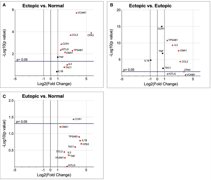
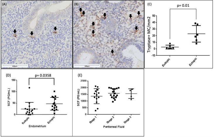
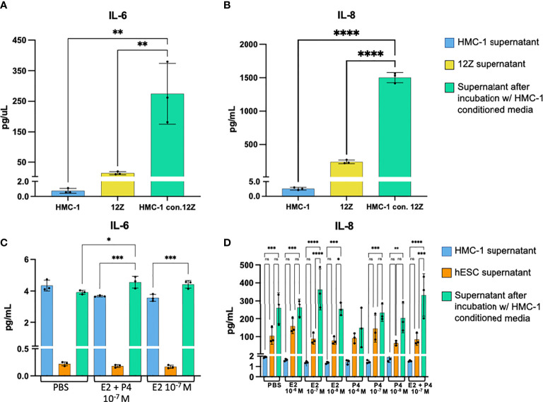
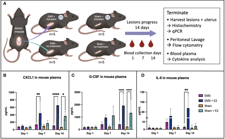
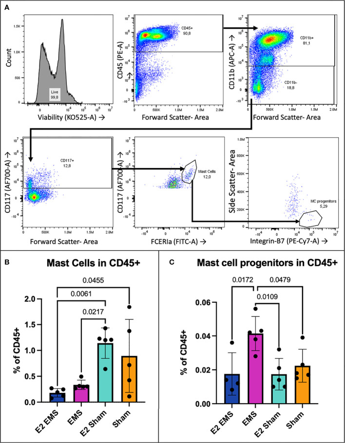
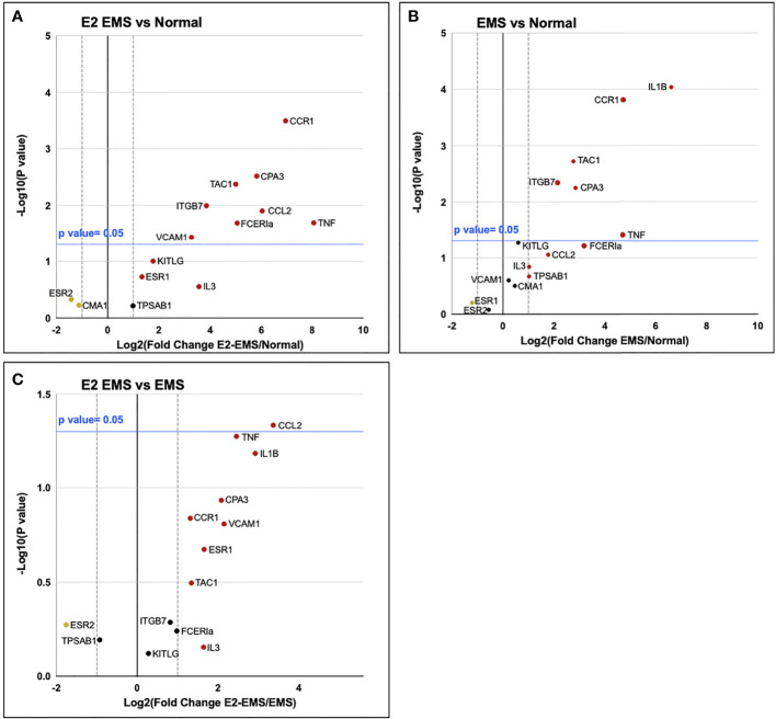
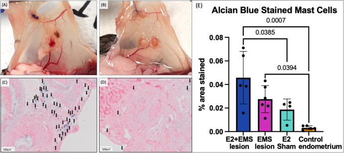
Endometriosis is an estrogen dependent, chronic inflammatory disease characterized by the growth of endometrial lining outside of the uterus. Mast cells have emerged as key players in regulating not only allergic responses but also other mechanisms such as angiogenesis, fibrosis, and pain. The influence of estrogen on mast cell function has also been recognized as a potential factor driving disease pathophysiology in number of allergic and chronic inflammatory conditions. However, precise information is lacking on the cross talk between endocrine and immune factors within the endometriotic lesions and whether that contributes to the involvement of mast cells with disease pathophysiology. In this study, we observed a significant increase in mast cell numbers within endometriotic lesions compared to matched eutopic endometrium from the same patients. Compared to eutopic endometrium, endometriotic lesions had significantly higher levels of stem cell factor (SCF), a potent growth factor critical for mast cell expansion, differentiation, and survival for tissue resident mast cells. Targeted mRNA Q-PCR array revealed that the endometriotic lesions harbour microenvironment (upregulation of CPA3, VCAM1, CCL2, CMA1, CCR1, and KITLG) that is conducive to mast cells recruitment and subsequent differentiation. To examine cross-talk of mast cells within the endometriotic lesion microenvironment, endometriotic epithelial cells (12Z) and endometrial stromal cells (hESC) incubated with mast cell-conditioned media showed significantly increased production of pro-inflammatory and chemokinetic cytokines. To further understand the impact of estrogen on mast cells in endometriosis, we induced endometriosis in C57BL/6 mice. Mature mast cells were significantly higher in peritoneal fluid of estrogen-treated mice compared to untreated mice within the sham operated groups. Mouse endometriotic lesion tissue revealed several genes (qRT-PCR) relevant in mast cell biology significantly upregulated in the estrogen treated, endometriosis-induced group compared to control endometrium. The endometriotic lesions from estrogen treated mice also had significantly higher density of Alcian blue stained mast cells compared to untreated lesions or control endometrium. Collectively, these findings suggest that endometriotic lesions provide a microenvironment necessary for recruitment and differentiation of mast cells. In turn, mast cells potentially release pro-inflammatory mediators that contribute to chronic pelvic pain and endometriosis disease progression.

Keywords: endometriosis; estrogenic inflammation; immune crosstalk; immune microenvironment; mast cell recruitment and maturation; stem cell factor.


Conflict of interest statement

The authors declare that the research was conducted in the absence of any commercial or financial relationships that could be construed as a potential conflict of interest.

Figures

Figure 1
Figure 1
Mast cell relevant genes are upregulated in endometriotic lesion tissue. Gene transcript fold change in ectopic endometriotic lesion (n=9) vs normal healthy endometrium (n=10) (A), eutopic endometrium (n=8) of endometriosis patients vs normal healthy endometrium (B), and ectopic endometriotic lesion vs eutopic endometrium of endometriosis patients (C). Vertical dotted lines represent a fold change of ±2, where data points outside this range have shown a fold change of more than 2.0. The blue horizontal line denotes p= 0.05 in -Log10, where data points above the blue line are significantly upregulated in the tested group.
Figure 2
Figure 2
Significantly higher mast cell numbers and stem cell factor concentrations found in ectopic endometriotic tissue than eutopic endometrium. Activated Mast cells are increased in human endometriotic lesions compared to matched eutopic endometrium. Immunohistochemistry for mast cell tryptase in eutopic endometrial samples (A) and endometriotic lesions (B) from stage III-IV endometriosis patients (n=6). Quantitative analysis (C) revealed significantly higher number of mast cells/mm2 in ectopic lesions compared to eutopic endometrium (p<0.01). Mast cell tryptase positive cells are indicated by black arrowheads. Scale bars: (A) 150µm, (B) 300µm, (C) 75µm, (D) 100µm. Stem cell factor concentration in human endometriotic lesions vs eutopic endometrium (n=15) (D), SCF concentrations in peritoneal fluid of endometriosis patients (n=14) stratified by disease stage (E).
Figure 3
Figure 3
Cytokine levels in 12Z and hESC supernatant without vs. with HMC-1 conditioned media. Concentration of IL-6 (A) and IL-8 (B) in 12Z supernatant without vs. with stimulation by HMC-1 conditioned media. Concentration of IL-6 (C) and IL-8 (D) in hESC supernatant without vs. with HMC-1 conditioned media in hormonal conditions. ****p= <0.0001, ***p= 0.0001—0.001, **p= 0.001—0.008, *p< 0.05.
Figure 4
Figure 4
Schematic diagram of syngeneic mouse model experiment used in this study (A). Plasma cytokine levels of CXCL1 (B), G-CSF (C) and IL-6 (D) were found to increase significantly in the estrogen treated EMS-induced group. *p<0.05, **p<0.0055, ****p<0.0001.
Figure 5
Figure 5
Mast cells in peritoneal fluid from mice induced with endometriosis. Mature mast cells were CD117+, FCERIa+, CD11b- (A). MCp were CD117+, FCERIa+, CD11b-, integrin β7+, SSClo (A). The estrogen treated sham operated group had significantly higher mast cell frequency within the CD45+ population compared to the untreated EMS group (p= 0.0256) (B). The E2 EMS group had significantly lower peritoneal mast cells than both E2 sham and untreated sham groups (p= 0.0057, 0.0414) (B). Frequency of MCp in the CD45+ population was significantly higher in EMS-induced mice vs. E2-treated EMS and E2-sham, untreated sham groups (p= 0.0172, 0.0109, 0.0479) (C).
Figure 6
Figure 6
Mast cell relevant genes are upregulated in endometriotic lesion tissue. Gene transcript fold change in estrogen-treated endometriotic lesion (n=5) vs normal healthy endometrium (n=5) (A), untreated endometriotic lesion (n=5) vs normal healthy endometrium (B), and estrogen-treated endometriotic lesion vs untreated endometriotic lesion (C) of endometriosis-induced and healthy control mice. Vertical dotted lines represent a fold change of ±2, where data points outside this range have shown a fold change of more than 2.0. The blue horizontal line denotes p= 0.05 in -Log10, where data points above the blue line are significantly upregulated in the tested group.
Figure 7
Figure 7
Murine endometriotic lesions, histochemistry, comparison of lesion MC presence to original uterine graft tissue. Visually notable qualitative size difference in estrogen-treated (A) vs untreated (B) mouse endometriotic lesions. Alcian blue staining of estrogen treated (C) and untreated (D) mouse endometriotic lesions, mast cells are denoted with black arrows. The % area stained with Alcian blue in lesions from E2-EMS mice (n= 5) and untreated EMS mice (n=6) was significantly higher compared to control endometrium (n= 5) (p= 0.0007, 0.0394) (E). Alcian blue staining was also significantly higher in E2-EMS lesions compared to endometrium of E2-sham mice (n=4) (p=0.0385). Scale bars: 100 μm (C, D).

References

    1. Giudice LC, Kao LC. Endometriosis. Lancet (2004) 364:1789–99. doi: 10.1016/S0140-6736(04)17403-5 - DOI - PubMed
    1. Vallvé-Juanico J, Houshdaran S, Giudice LC. The endometrial immune environment of women with endometriosis. Hum Reprod Update (2019) 25:565–92. doi: 10.1093/humupd/dmz018 - DOI - PMC - PubMed
    1. Kirchhoff D, Kaulfuss S, Fuhrmann U, Maurer M, Zollner TM. Mast cells in endometriosis: Guilty or innocent bystanders? Expert Opin Ther Targets (2012) 16:237–41. doi: 10.1517/14728222.2012.661415 - DOI - PubMed
    1. Galli SJ. Mast cells and basophils, 1st edition. Curr Opin Hematol (2000) 707:. doi: 10.1097/00062752-200001000-00007 - DOI - PubMed
    1. Sugamata M, Ihara T, Uchiide I. Increase of activated mast cells in human endometriosis. Am J Reprod Immunol (2005) 53:120–5. doi: 10.1111/j.1600-0897.2005.00254.x - DOI - PubMed

Publication types