. 2022 Aug 13:21:44-56.
doi: 10.1016/j.bioactmat.2022.07.028. eCollection 2023 Mar.

Simvastatin-hydroxyapatite coatings prevent biofilm formation and improve bone formation in implant-associated infections

Affiliations

Simvastatin-hydroxyapatite coatings prevent biofilm formation and improve bone formation in implant-associated infections

Tiantong Sun et al. Bioact Mater. .

Abstract

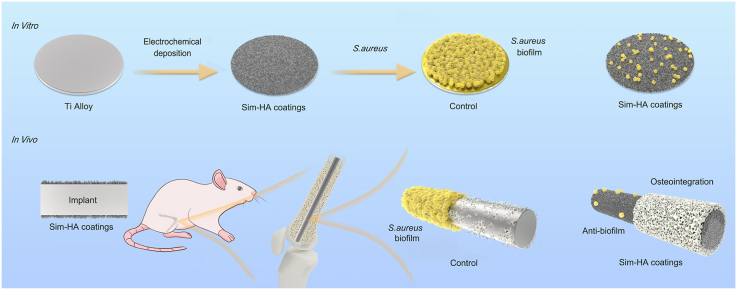
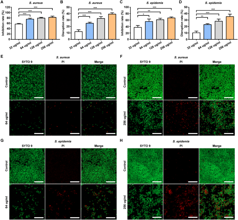
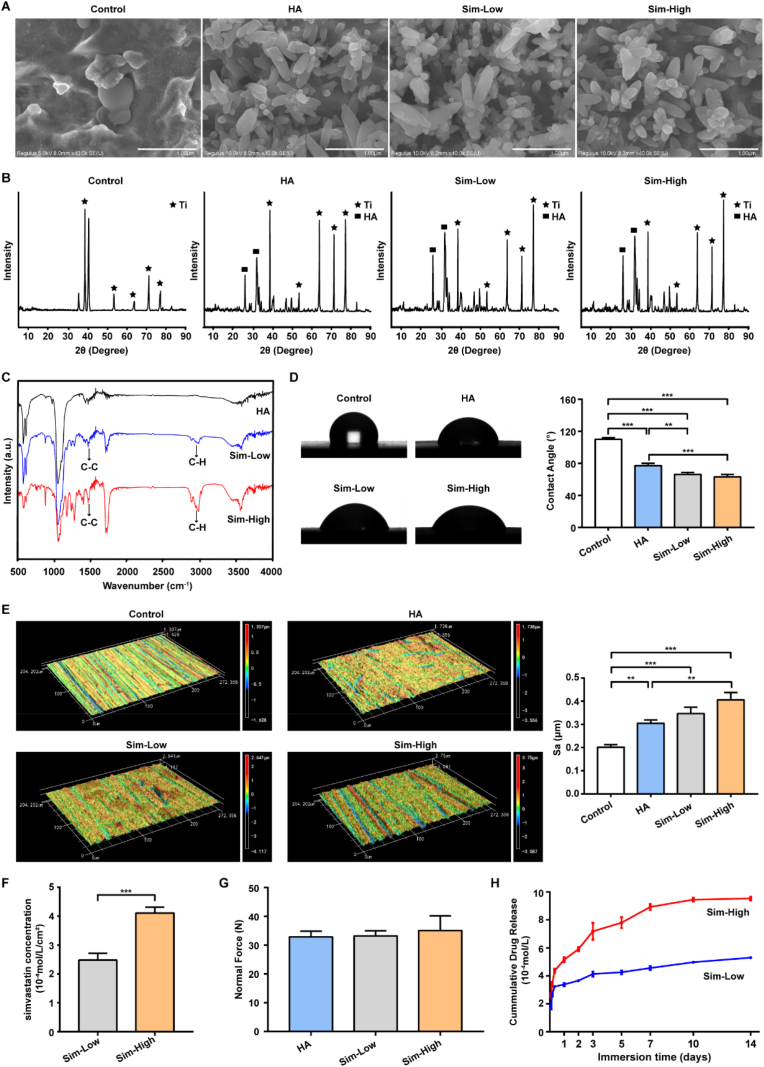
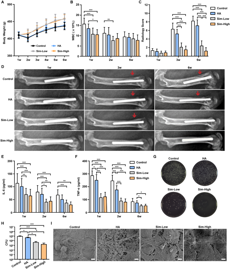
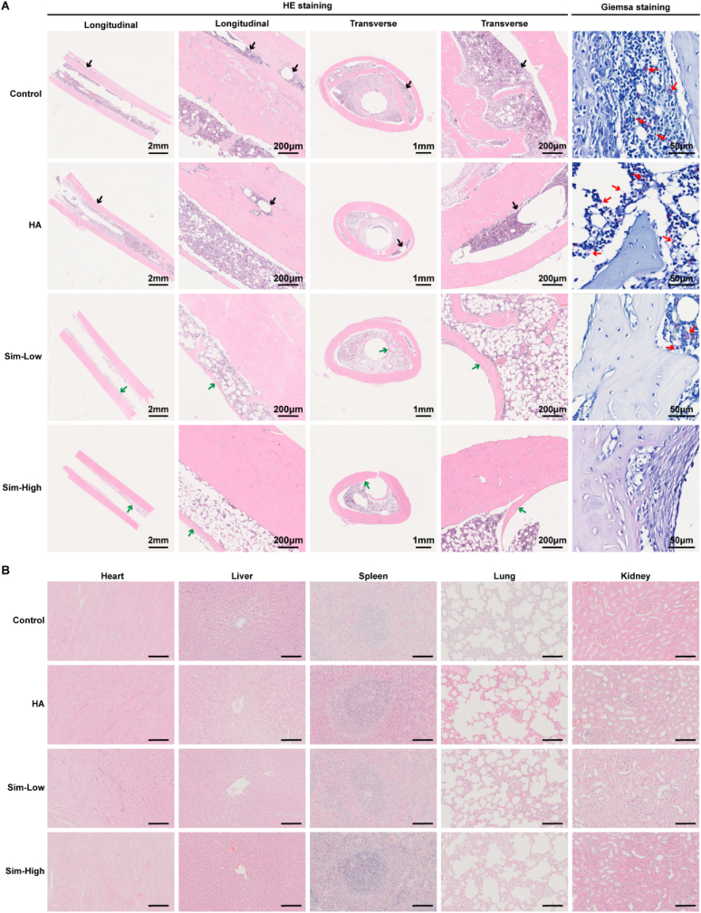
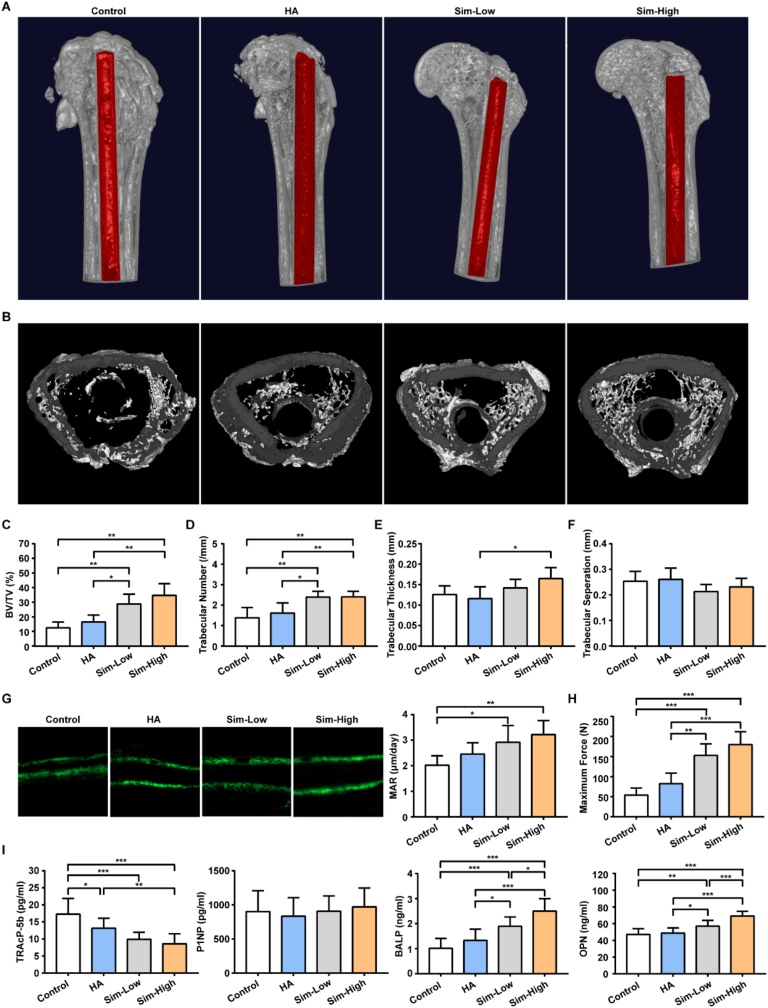
Implant-associated infections (IAIs) caused by biofilm formation are the most devastating complications of orthopedic surgery. Statins have been commonly and safely used drugs for hypercholesterolemia for many years. Here, we report that simvastatin-hydroxyapatite-coated titanium alloy prevents biofilm-associated infections. The antibacterial properties of simvastatin against Staphylococcus aureus and Staphylococcus epidermidis biofilms in vitro was confirmed by crystal violet staining and live-dead bacterial staining. We developed a simvastatin-and hydroxyapatite (Sim-HA)-coated titanium alloy via electrochemical deposition. Sim-HA coatings inhibited Staphylococcus aureus biofilm formation and improved the biocompatibility of the titanium alloy. Sim-HA coatings effectively prevented Staphylococcus aureus IAI in rat femurs, as confirmed by radiological assessment and histological examination. The antibacterial effects of the Sim-HA coatings were attributed to their inhibitory effects on biofilm formation, as verified by scanning electron microscopic observations and bacterial spread plate analysis. In addition, the Sim-HA coatings enhanced osteogenesis and osteointegration, as verified by micro-CT, histological evaluation, and biomechanical pull-out tests. In summary, Sim-HA coatings are promising implant materials for protection against biofilm-associated infections.

Keywords: Antibacterial; Biofilm; Implant-associated infections; Osteogenesis; Simvastatin.


Conflict of interest statement

None.

Figures

Image 1
Graphical abstract
Fig. 1
Fig. 1
Effects of simvastatin on biofilm inhibition and disruption. A-DS. aureus and S. epidermis were treated with simvastatin at different concentrations for 24 h. The inhibition effect and disruption effect were measured by crystal violet staining. Data are shown as the mean ± SD, n = 3, * for P < 0.05, ** for P < 0.01, *** for P < 0.001. E-H Confocal fluorescence images showed S. aureus and S. epidermis biofilms after 24 h of treatment with simvastatin at different concentrations. Live cells were stained green and dead cells were stained red. Scale bar: 20 μm.
Fig. 2
Fig. 2
Characterization and properties of Sim-HA-Ti6Al4V. A SEM micrograph of uncoated or coated Ti6Al4V with HA and different concentrations of simvastatin. Scale bar: 1 μm. B XRD patterns of HA precipitation and Ti precipitation. C FTIR spectra of Ti6Al4V coated with HA and different concentrations of simvastatin. D Representative images and statistics of contact angles in four groups. E Representative topographical images of surfaces determined by 3D laser microscopy and statistics of surface area roughness (Sa) in four groups. F Concentration analysis of simvastatin in two Sim-HA coatings by LC-MS/MS. G Bonding strength (Normal Force) of HA and Sim-HA coatings. H The cumulative drug release profiles for two Sim-HA coatings. Data are shown as the mean ± SD, n = 3, ** for P < 0.01, *** for P < 0.001.
Fig. 3
Fig. 3
Antibacterial effect and biocompatibility of Sim-HA-Ti6Al4V. A Confocal fluorescence images of Ti6Al4V, Ti6Al4V-HA and Sim-HA-Ti6Al4V for S. aureus biofilm formation by laser confocal microscopy, scale bar: 50 μm. B SEM images of four implants for S. aureus biofilm formation, scale bar: 2 μm. C Images and statistics of spread plate assay. D Rat BMSCs adhesion on different four implants, scale bar: 50 μm. E Proliferation rate of rat BMSCs grown on the four different implants. F ALP activity of BMSCs on four different implants. Data are shown as the mean ± SD, n = 3. * for P < 0.05. ** for P < 0.01, *** for P < 0.001.
Fig. 4
Fig. 4
Antibacterial evaluation of Sim-HA-Ti6Al4V in vivo. A Changes in the body weights of the rats in the four groups during the 6 weeks after surgery. B Changes in rat blood WBCs in the four groups at 1 w, 2 w, and 6 w after surgery. C Radiology scores of X-rays of four groups at 1 w, 3 w, and 6 w after surgery (n = 10). D Representative images of rat femur at 1 w, 3 w, and 6 w after surgery. The red arrows indicate destruction of the cortical bone of the distal femur. E-F Plasma concentrations of IL-6 and TNF-α in rats in the four groups at 1 w, 2 w, and 6 w after surgery. G Culture of bacteria released from four different implants. H Quantitative analysis of released bacteria from four different implants (n = 3). I SEM images of four different implants 6 w after surgery, scale bar:2 μm. Data are shown as the mean ± SD, n = 12. *for P < 0.05, ** for P < 0.01, *** for P < 0.001.
Fig. 5
Fig. 5
Histological evaluation of the Sim-HA-Ti6Al4V in vivo. A Representative histological images of hematoxylin and eosin staining and Giemsa staining of rat femurs. The black arrows represent massive destruction of cortical bone. The green arrows indicate new bone formation around implants. The red arrows indicate bacteria remaining in the bone tissue. B Hematoxylin and eosin staining analyses of different organs, including the heart, liver, spleen, lung, and kidney. n = 3.
Fig. 6
Fig. 6
Osteogenesis evaluation of the Sim-HA-Ti6Al4V in vivo. A The 3D reconstruction images of rat distal femur 6 w after surgery, implants are marked in red. B Cross-section images of rat distal femur metaphysis. CF Quantitative analysis of the trabecular microarchitecture of the distal femur metaphysis 6 w after surgery (n = 6). G Representative images of calcein double-labeling and MAR in the rat distal femurs of the four groups (n = 6). H Maximum force of biomechanical pull-out test in the four groups. (n = 5) I Plasma concentrations of TRAcP-5b, P1NP, BALP and OPN in the four groups 6 w after surgery (n = 10). Data are shown as the mean ± SD. * for P < 0.05, ** for P < 0.01, *** for P < 0.001.

Similar articles

Cited by

References

    1. Zimmerli W. Clinical presentation and treatment of orthopaedic implant-associated infection. J. Intern. Med. 2014;276(2):111–119. - PubMed
    1. Kapadia B.H., Berg R.A., Daley J.A., Fritz J., Bhave A., Mont M.A. Periprosthetic joint infection. Lancet. 2016;387(10016):386–394. - PubMed
    1. Ashbaugh A.G., Jiang X., Zheng J., Tsai A.S., Kim W.S., Thompson J.M., et al. Polymeric nanofiber coating with tunable combinatorial antibiotic delivery prevents biofilm-associated infection in vivo. Proc. Natl. Acad. Sci. U. S. A. 2016;113(45):E6919–e6928. - PMC - PubMed
    1. Vallet-Regi M., Lozano D., Gonzalez B., Izquierdo-Barba I. Biomaterials against bone infection. Adv. Healthc. Mater. 2020;9(13) - PMC - PubMed
    1. Zimmerli W., Sendi P. Orthopaedic biofilm infections. Apmis. 2017;125(4):353–364. - PubMed

LinkOut - more resources