Review
. 2022 Aug 9:9:968752.
doi: 10.3389/fcvm.2022.968752. eCollection 2022.

Advances in cell death mechanisms involved in viral myocarditis

Affiliations
Review

Advances in cell death mechanisms involved in viral myocarditis

Yang Yang et al. Front Cardiovasc Med. .

Abstract

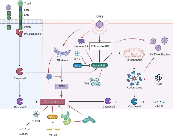
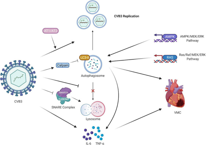
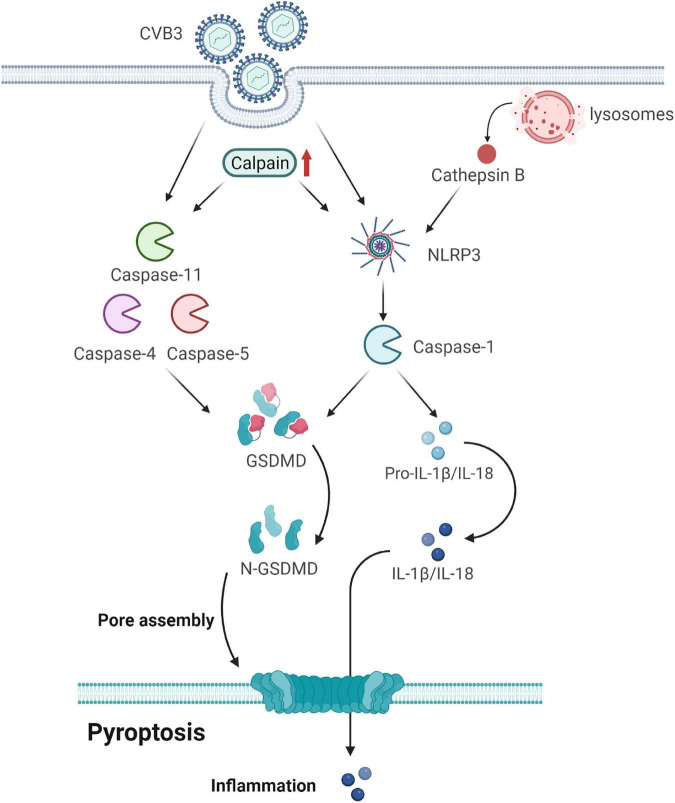
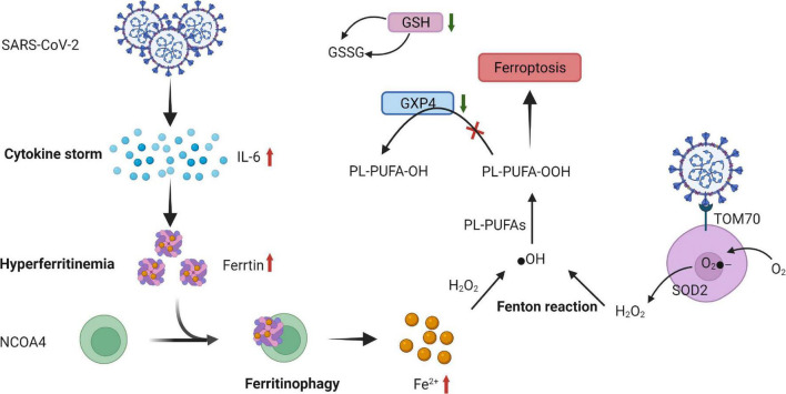
Viral myocarditis is an acute inflammatory disease of the myocardium. Although many etiopathogenic factors exist, coxsackievirus B3 is a the leading cause of viral myocarditis. Abnormal cardiomyocyte death is the underlying problem for most cardiovascular diseases and fatalities. Various types of cell death occur and are regulated to varying degrees. In this review, we discuss the different cell death mechanisms in viral myocarditis and the potential interactions between them. We also explore the role and mechanism of cardiomyocyte death with severe acute respiratory syndrome coronavirus 2 (SARS-CoV-2) infection. Exploring the mechanisms may help in the early identification and the development of effective treatments, thus improving the quality of life of patients with viral myocarditis. We believe that the inhibition of cardiomyocyte death has immense therapeutic potential in increasing the longevity and health of the heart.

Keywords: CVB3; SARS-CoV-2; VMC; apoptosis; autophagy; ferroptosis; necrosis; pyroptosis.


Conflict of interest statement

The authors declare that the research was conducted in the absence of any commercial or financial relationships that could be construed as a potential conflict of interest.

Figures

FIGURE 1
FIGURE 1
Coxsackievirus B3 (CVB3) manipulates cell apoptosis. CVB3 infection can markedly induce myocardial apoptosis via death receptor–mediated and mitochondrial-mediated signaling pathways. Viral infection induces abnormal expression of Fas antigen in the myocardium and cross-links with FasL of active cells. Activated CTL binds to target cells via Fas/FasL and causes apoptosis. The PI3K and mTOR-signaling pathways participate in the CVB3-induced VMC by mediating the proapoptosis factor Bim, Bax, caspase-9, caspase-3, and viral replication. The inhibition of HDAC activity increases CVB3 replication by enhancing autophagosome formation and ensuing increased myocardial apoptosis. IL-17A mediated cardiomyocyte apoptosis by regulating the Bax/Bcl-2 ratio. miR-133 targets caspase-9 and promotes cardiac cell apoptosis. miR-15 could suppress cell viability and promote CVB3-induced cell apoptosis by modulating the NLRX1-mediated NLRP3 inflammasome. miR-21 alleviates CVB3-induced myocarditis by protecting myocardial apoptosis and repressing PDCD4 expression. CVB3-stimulated cytotoxicity can be inhibited by kinase ERK5, coupled with p38 kinase activity. However, p38 promotes apoptosis through ERK1/2 inhibition indirectly. c-Fos can compose AP-1 with c-jun gene products that regulate the transcription of apoptosis-related genes. Picornavirus protease 2A in CVB3 induced apoptosis through multiple converging pathways. ER-initiated apoptosis was induced by CVB3-infected cardiomyocytes and caused myocardial apoptosis through ER stress by the PERK pathway. CP, which is located within the endoplasmic reticulum Ca2+ binding proteins, can relieve ERS-initiated apoptosis in VCM. CVB3, coxsackievirus B3; CTL, cytotoxic T lymphocytes; ER, endoplasmic reticulum; Bcl-2, B-cell lymphoma 2; Bax, Bcl-2-associated X protein; HDAC, histone deacetylase; NLRP3, NLR family pyrin domain containing 3; PDCD4, programmed cell death 4; ER, endoplasmic reticulum; CP, calumenin protein.
FIGURE 2
FIGURE 2
Regulation of CVB3 and autophagy in VMC. CVB3 infection triggers the formation of autophagosomes and uses the autophagosomal pathway for replication. CVB3 infection in cardiomyocytes activated calpain and increased. The inhibition of calpain activity led to the accumulation of LC3-II protein expression, impairing the autophagic flux. AT1 and AT2 regulate cardiomyocyte autophagy activity by propagating viral replication, thus triggering autophagic cell death. CVB3 might directly or indirectly induce autophagy via the AMPK/MEK/ERK and Ras/Raf/MEK/ERK signaling pathways. CVB3 inhibits the fusion of lysosomes with autophagosomes. CVB3 specifically targets the SNARE protein SNAP29 and the adaptor protein PLEKHM1, both of which regulate autophagosome fusion, for cleavage through the catalytic activity of viral proteinase 3C; this process ultimately impairs the formation of SNARE complexes. The release of proinflammatory cytokines also participates in the cardiac fibroblasts caused by CVB3 infection, and the downregulation of autophagy suppresses them. CVB3, coxsackievirus B3; VMC, viral myocarditis; LC3, light chain 3; AT1 and AT2, Type I and II angiotensin II receptors.
FIGURE 3
FIGURE 3
Gasdermin D (GSDMD) forms membrane pores to cause pyroptosis. CVB3 infection initiates pyroptosis by canonical caspase-1-dependent and non-canonical caspase-4/5/11-mediated pyroptosis pathways. In the canonical pyroptosis pathway, cells recognize intracellular pathogens through many PRRs and form NLRP3, which activates caspase-1. Caspase-1 processes and activates IL-1β and IL-18; it also cleaves GSDMD to release the membrane pore-forming GSDMD-N domain. GSDMD-N pores promote the release of activated IL-1β and IL-18. In the non-canonical pyroptosis pathway, cytosolic LPS binds to caspase-4/5/11 and triggers the cleavage of GSDMD but not of IL-1β and IL-18. Calpain is activated after CVB3 infection, accompanied by an increase in pyroptosis by promoting the canonical NLRP3 inflammasome/caspase-1-mediated and non-canonical caspase-11-mediated pyroptosis pathways. CVB3 infection damages the lysosomes and releases the lysosomal contents, including cathepsin B, into the cytosol. Cathepsin B exaggerates VMC by regulating the activation of NLRP3. GSDMD, gasdermin D; CVB3, coxsackievirus B3; PRRs, pattern-recognition receptors; LPS, lipopolysaccharide; VMC, viral myocarditis.
FIGURE 4
FIGURE 4
Proposed mechanism of ferroptosis in SARS-CoV-2 infection. SARS-CoV-2-related increase in cytokines, especially IL-6 causing hyperferritinemia, is characterized by the increase in intracellular iron and ferritin. This increased ferritin binds to NCOA4 and is delivered to autophagosomes, causing ferritinophagy and triggering an increase in the labile iron pool, which induces OH through the Fenton reaction and, eventually, through PL-PUFA peroxidation, which promotes ferroptosis. The expression of GSH and ferroptosis-associated GPX4 is suppressed by severe acute respiratory syndrome coronavirus 2 (SARS-CoV-2) infection. Moreover, a low GSH pool and downregulation of GPX4 gene expression caused by SARS-CoV-2 infection facilitate ferroptosis. Orf9b, one of the accessory proteins of SARS-CoV-2, increases ROS generation by binding to TOM70 at the surface of the mitochondria membrane. O2- is produced by ETC on the internal membrane of the mitochondria and then converted to further H2O2 by SOD and eventually, by Fenton reaction, transformed into ⋅OH, triggering LOOH generation from PUFAs that promotes ferroptosis. SARS-CoV-2, severe acute respiratory syndrome coronavirus 2; IL-6, interleukin-6; NCOA4, nuclear receptor coactivator 4; ⋅OH, hydroxyl radical; PL-PUFAs, phospholipid polyunsaturated fatty acids; PUFA-OH, phospholipid polyunsaturated fatty acid alcohols; PL-PUFA-OOH, phospholipid polyunsaturated fatty acid peroxides; GSH, glutathione; GSSG, oxidized glutathione; GPX4, glutathione peroxidase; Orf9B, open reading frame-9b; ROS, reactive oxygen species; TOM70, translocase of outer membrane 70; O2-, superoxide; ETC, electron transport chain; H2O2, hydrogen peroxide; SOD, superoxide dismutase; LOOH, peroxides.
FIGURE 5
FIGURE 5
The main characteristics of histology in VMC. (A) Active myocarditis is characterized by an inflammatory cellular infiltrate with numerous necrotic myocytes. (B) Representative histopathology in a borderline myocarditis group. The inflammatory region is showed with several large foci of cellular infiltrations. (C) Diffuse lymphocytic infiltration of myocardium is described as lymphocytic, eosinophilic, or granulomatous in endomyocardial biopsy specimens. VMC, viral myocarditis.

References

    1. Dhakal BP, Sweitzer NK, Indik JH, Acharya D, William P. SARS-CoV-2 infection and cardiovascular disease: COVID-19 heart. Heart Lung Circ. (2020) 29:973–87. 10.1016/j.hlc.2020.05.101 - DOI - PMC - PubMed
    1. Zhou L, He X, Gao B, Xiong S. Inhibition of histone deacetylase activity aggravates coxsackievirus B3-induced myocarditis by promoting viral replication and myocardial apoptosis. J Virol. (2015) 89:10512–23. 10.1128/JVI.01028-15 - DOI - PMC - PubMed
    1. Liao YH, Xia N, Zhou SF, Tang TT, Yan XX, Lv BJ, et al. Interleukin-17A contributes to myocardial ischemia/reperfusion injury by regulating cardiomyocyte apoptosis and neutrophil infiltration. J Am Coll Cardiol. (2012) 59:420–9. 10.1016/j.jacc.2011.10.863 - DOI - PMC - PubMed
    1. Lv S, Rong J, Ren S, Wu M, Li M, Zhu Y, et al. Epidemiology and diagnosis of viral myocarditis. Hellenic J Cardiol. (2013) 54:382–91. - PubMed
    1. Esfandiarei M, McManus BM. Molecular biology and pathogenesis of viral myocarditis. Ann Rev Pathol. (2008) 3:127–55. 10.1146/annurev.pathmechdis.3.121806.151534 - DOI - PubMed