. 2022 Aug 16:2022:8360481.
doi: 10.1155/2022/8360481. eCollection 2022.

Prognostic Values of BolA Family Member Expression in Hepatocellular Carcinoma

Affiliations

Prognostic Values of BolA Family Member Expression in Hepatocellular Carcinoma

Dong Wang et al. Biomed Res Int. .

Abstract

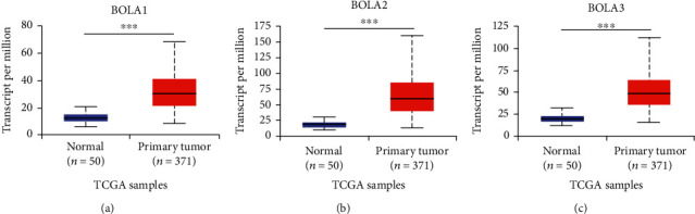
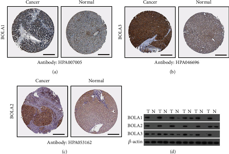
The BolA gene family member (BOLA1-3) plays an important role in regulating normal and pathological biological processes including liver tumorigenesis. However, their expression patterns as prognostic factors in hepatocellular carcinoma (HCC) patients have not to be elucidated. We examined the transcriptional expressions and survival data of BolA family member in patients with HCC from online databases including ONCOMINE, TCGA, UALCAN, Gene Expression Profiling Interactive Analysis (GEPIA), Kaplan-Meier plotter, SurvExpress, cBioPortal, and Exobase. Network molecular interaction views of BolA family members and their neighborhoods were constructed by the IntAct web server. In our research, we had found that the expression levels of BolA /2/3 mRNA were higher in HCC tissue than in normal liver tissues from TGCA databases. Moreover, the BolA family gene expression level is significantly associated with distinct tumor pathological grade, TMN stage, and overall survival (OS). The BolA family can be considered as prognostic risk biomarkers of HCC. A small number of BolA gene-mutated samples were detected in the HCC tissue. IntAct analysis revealed that BolA1/2/3 was closely associated with the GLRX3 expression in HCC, which is implicated in the regulation of the cellular iron homeostasis and tumor growth. Furthermore, prognostic values of altered BolAs and their neighbor GLRX3 gene in HCC patients were validated by SurvExpress analysis. In conclusion, the membrane BolA family identified in this study provides very useful information for the mechanism of hepatic tumorigenesis.


Conflict of interest statement

The authors declare that they have no potential competing interests in this work.

Figures

Figure 1
Figure 1
Transcriptional expression of BolA family members in 20 different types of cancer types (ONCOMINE database). Notes: the BOLA1\2\3 mRNA expression (cancer tissue vs. normal tissue) was compared by Students' t-test. Cut-off of change values was as follows: P value: 0.01, fold change: 1.5, gene rank: 10%, and data type: mRNA.
Figure 2
Figure 2
The BOLA1\2\3 mRNA expressions in HCC and adjacent nontumorous tissues (UALCAN database). Notes: BolA family gene mRNA was higher in HCC tissues compared to nontumorous tissues. Statistically significant changes were indicated with asterisks. ∗∗∗P < 0.001.
Figure 3
Figure 3
Protein expressions of BolA family members in human normal liver tissue and HCC (Human Protein Atlas database). Notes: BOLA1/2/3 proteins were lower in normal liver tissues than in HCC tissues. BOLA1 antibody HPA007005, BOLA2 antibody HPA046696, and BOLA3 antibody HPA053162. Scale bar is 100 μm (a)–(c). (d) The BOLA1/2/3 expression in the HCC and nontumor samples was as follows, and we had found that the BOLA1/2/3 BOLA1\2\3 expression level was higher in HCC than in the nontumor. T: tumor; NT: nontumor.
Figure 4
Figure 4
BolA family member mRNA expressions with clinicopathologic parameters in HCC (UALCAN database). Notes: mRNA expressions of BolA family members were significantly related to tumor grades, and as tumor grade increased, the mRAN expressions of BolAs tended to be higher (a). mRNA expressions of BolA family members were remarkably correlated with clinical stages, and patients who were in more advanced stages tended to express higher mRNA expression of BolAs (b). Data are mean ± SEM. P < 0.05; ∗∗P < 0.01; ∗∗∗P < 0.001.
Figure 5
Figure 5
The BolA family members with clinical outcomes in HCC patients by Kaplan-Meier curves (GEPIA database and SurvExpress database). Notes: overall survival data of BolA family members are generated from the GEPIA web server (a)–(c). Prognostic risk of the mRNA expression of BolA family members in HCC patients (d). The concordance index and P value of log-rank testing equality of survival curves are indicated. The box plots indicate the difference in the expression of gene between risks groups, and P values are derived from t-test between both groups (e).
Figure 6
Figure 6
Network protein-protein interaction views of BolA family members and their neighborhood in TCGA-LIHC patients (IntAct database). Notes: network of molecular interaction was constructed by IntAct. Red marker indicates activation relationship of two intermediate related genes, including BIRC2, XIAP, C1orf94, HDX, BIRC7, BCKDHA, PMPCA, PMPCB, GLRX5, and SDHAF3. Purple marker indicates activation relationship of three intermediate related genes, including GLRX3 and DDIT4L.
Figure 7
Figure 7
Prognostic values of BolA family altered neighbor genes in HCC patients (Kaplan-Meier plotter). Notes: representative altered neighbor genes of BolA family members, including GLRX3 (a), BIRC7 (b), BIRC2(c), BCKDHA (d), PMPC8 (e), and GLRX5 (f).
Figure 8
Figure 8
Combinatory BolA family members and GLRX3 and PMPCB predict survival of TCGA-LIHC patients (SurvExpress dataset). Notes: (a) Kaplan–Meier survival curve of 361 TCGA HCC samples using the SurvExpress database, based on the low or high risk for a poor outcome. (b) The high expression of five hub genes is correlated with high risk, poor prognosis, and shorter overall survival time. High- and low-risk groups are labeled with red and green curves, respectively. The box plots indicate the difference in expression of gene between risks groups, and P values are derived from t-test between both groups.

References

    1. Siegel R. L., Miller K. D., Jemal A. Cancer statistics. CA: a Cancer Journal for Clinicians . 2019;69(1):7–34. doi: 10.3322/caac.21551. - DOI - PubMed
    1. Ba Q., Hao M., Huang H., et al. Iron deprivation suppresses hepatocellular carcinoma growth in experimental studies. Clinical Cancer Research . 2011;17(24):7625–7633. doi: 10.1158/1078-0432.CCR-10-3099. - DOI - PubMed
    1. Torti S. V., Torti F. M. Iron and cancer: more ore to be mined. Nature Reviews. Cancer . 2013;13(5):342–355. doi: 10.1038/nrc3495. - DOI - PMC - PubMed
    1. Zhou Y. B., Cao J. B., Wan B. B., et al. hBolA, novel non-classical secreted proteins, belonging to different BolA family with functional divergence. Molecular and Cellular Biochemistry . 2008;317(1-2):61–68. doi: 10.1007/s11010-008-9809-2. - DOI - PubMed
    1. Uzarska M. A., Nasta V., Weiler B. D., et al. Mitochondrial Bol1 and Bol3 function as assembly factors for specific iron-sulfur proteins. eLife . 2016;5 doi: 10.7554/eLife.16673. - DOI - PMC - PubMed