. 2022 Sep 21;33(9):1685-1697.
doi: 10.1021/acs.bioconjchem.2c00305. Epub 2022 Aug 26.

Porous Silicon Nanoparticles Targeted to the Extracellular Matrix for Therapeutic Protein Delivery in Traumatic Brain Injury

Affiliations

Porous Silicon Nanoparticles Targeted to the Extracellular Matrix for Therapeutic Protein Delivery in Traumatic Brain Injury

Lauren E Waggoner et al. Bioconjug Chem. .

Abstract

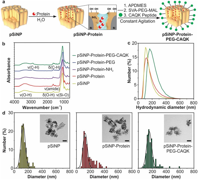
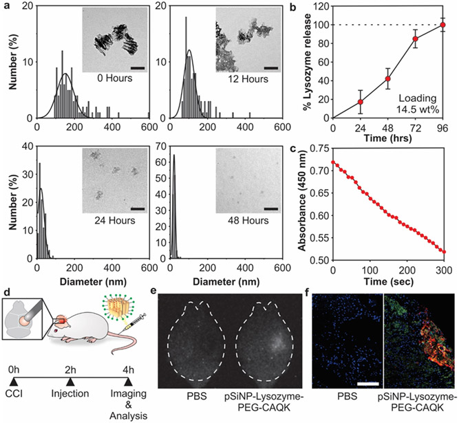
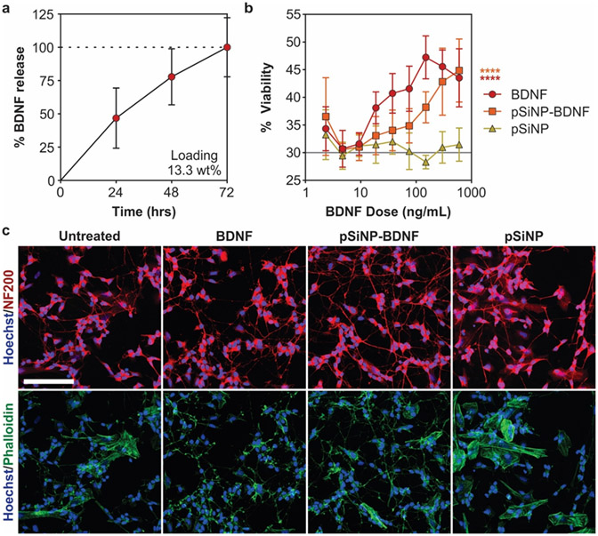
Traumatic brain injury (TBI) is a major cause of disability and death among children and young adults in the United States, yet there are currently no treatments that improve the long-term brain health of patients. One promising therapeutic for TBI is brain-derived neurotrophic factor (BDNF), a protein that promotes neurogenesis and neuron survival. However, outstanding challenges to the systemic delivery of BDNF are its instability in blood, poor transport into the brain, and short half-life in circulation and brain tissue. Here, BDNF is encapsulated into an engineered, biodegradable porous silicon nanoparticle (pSiNP) in order to deliver bioactive BDNF to injured brain tissue after TBI. The pSiNP carrier is modified with the targeting ligand CAQK, a peptide that binds to extracellular matrix components upregulated after TBI. The protein cargo retains bioactivity after release from the pSiNP carrier, and systemic administration of the CAQK-modified pSiNPs results in effective delivery of the protein cargo to injured brain regions in a mouse model of TBI. When administered after injury, the CAQK-targeted pSiNP delivery system for BDNF reduces lesion volumes compared to free BDNF, supporting the hypothesis that pSiNPs mediate therapeutic protein delivery after systemic administration to improve outcomes in TBI.


Conflict of interest statement

CONFLICT DISCLOSURE

MJS is a scientific founder (SF), member of the Board of Directors (BOD), Advisory Board (AB), Scientific Advisory Board (SAB), acts as a paid consultant (PC) or has an equity interest (EI) in the following: Aivocode, Inc (AB, EI); Bejing ITEC Technologies (SAB, PC); Cend Therapeutics (SF, BOD, EI); Illumina (EI); Matrix Technologies (EI); NanoVision Bio (SAB, EI); Pacific Integrated Energy (AB, EI); Quanterix (EI); Spinnaker Biosciences, Inc. (SF, BOD, EI); TruTag Technologies (SAB, EI); and Well-Healthcare Technologies (SAB, PC). MJS is also a Guest Professor at Zhejiang University, China. Although one or more of the grants that supported this research has been identified for conflict of interest management based on the overall scope of the project and its potential benefit to the companies listed, the research findings included in this publication may not necessarily relate to their interests. The terms of these arrangements have been reviewed and approved by the University of California, San Diego in accordance with its conflict of interest policies.

Figures

Figure 1:
Figure 1:. Synthesis and characterization of CAQK-targeted pSiNPs encapsulating a protein cargo.
(a) Schematic of protein loading, surface modification, and CAQK-functionalization of pSiNPs. (b) FTIR analysis of pSiNPs during synthesis steps. Nanoparticle diameters as measured by (c) DLS and (d) TEM (scale bar in inset = 100 nm).
Figure 2:
Figure 2:. Loading and biodistribution of model protein in pSiNPs.
(a) Analysis of pSiNP-Lysozyme-PEG-CAQK size by TEM image analysis after degradation in PBS at 37 °C after 0, 12, 24, and 48 hours (scale bar = 100 nm). (b) Time-dependent release of model protein lysozyme from pSiNPs in PBS at 37 °C, measured by BCA assay. The mass percentage loading of lysozyme in the pSiNP constructs was 14.5% by mass relative to the pSiNP-protein construct. (c) Activity of lysozyme after release from pSiNPs. Lysozyme activity was assayed through the hydrolysis of Micrococcus lysodeikticus, measured by loss of absorbance at 450 nm. (d) Schematic depiction of the protocol followed in the biodistribution study. The right hemisphere was injured, followed by intravenous administration of pSiNP-Lysozyme-PEG-CAQK 2 hours post-CCI, and brains collection for downstream imaging and analysis 2 hours post-injection. (e) Time-gated image of pSiNPs in whole brains and (f) confocal image of lysozyme model protein (red), CAQK (green), and nuclei (blue) from injured brain sections (scale bar = 200 μm).
Figure 3:
Figure 3:. BDNF loading and activity in differentiated SH-SY5Y cultures.
(a) Time-dependent release of BDNF from pSiNPs in PBS buffer at 37 °C, quantified by ELISA. The loading of BDNF in the pSiNPs was 13.3% by mass relative to the pSiNP-protein construct. (b) Cell viability in retinoic acid-differentiated SH-SY5Y cultures treated with BDNF, pSiNP, and pSiNP-BDNF. The “pSiNP" control trace corresponds to a concentration of pSiNPs that is the same amount of Si by mass as was used in the pSiNP-BDNF formulation; i.e., each point in the pSiNP control trace corresponds to a mass/volume of empty pSiNPs that is ~7.5x the ng/mL value indicated on the x-axis. (Gray line represents untreated cells; n=6, mean ± SD, **** p ≤ 0.0001 Two-way ANOVA with Dunnett's post-test compared to pSiNP control). (c) Representative images of SH-SY5Y cells treated for 72 hours with matched concentrations of 300 ng/mL BDNF and stained with NF200 (red), phalloidin (green), and Hoechst (blue) (scale bar =100 μm).
Figure 4:
Figure 4:. Lesion volume decreases after pSiNP-BDNF treatment.
(a) Schematic and timeline of injury, treatment, and lesion volume analysis. (b) Changes in lesion volumes relative to PBS-treated controls. (n = 7 per group, mean ± SEM, # p = 0.13 One-way ANOVA with Dunnett's post-test compared to PBS control). Both pSiNP and pSiNP-BDNF formulations contained PEG-CAQK surface chemistry as described in the text. (c) Representative images of H&E-stained coronal brain sections at 1.5 mm caudal from bregma for each treatment group with measured lesion area filled in gray (scale bar = 1 mm).

Similar articles

Cited by

References

    1. Surveillance Report of Traumatic Brain Injury-Related Emergency Department Visits, Hospitalizations, and Deaths. - PMC - PubMed
    1. Dean PJA; Sterr A Long-Term Effects of Mild Traumatic Brain Injury on Cognitive Performance. Front. Hum. Neurosci 2013, 7. 10.3389/fnhum.2013.00030. - DOI - PMC - PubMed
    1. Vanderploeg RD; Curtiss G; Belanger HG Long-Term Neuropsychological Outcomes Following Mild Traumatic Brain Injury. J. Int. Neuropsychol. Soc 2005, 11 (3), 228–236. 10.1017/S1355617705050289. - DOI - PubMed
    1. Werner C; Engelhard K Pathophysiology of Traumatic Brain Injury. Br. J. Anaesth 2007, 99 (1), 4–9. 10.1093/bja/aem131. - DOI - PubMed
    1. Nagahara AH; Tuszynski MH Potential Therapeutic Uses of BDNF in Neurological and Psychiatric Disorders. Nat. Rev. Drug Discov 2011, 10 (3), 209–219. 10.1038/nrd3366. - DOI - PubMed

Publication types