. 2023 Mar;18(3):560-567.
doi: 10.4103/1673-5374.346551.

Exosomal miR-23b from bone marrow mesenchymal stem cells alleviates oxidative stress and pyroptosis after intracerebral hemorrhage

Affiliations

Exosomal miR-23b from bone marrow mesenchymal stem cells alleviates oxidative stress and pyroptosis after intracerebral hemorrhage

Liu-Ting Hu et al. Neural Regen Res. 2023 Mar.

Abstract

Our previous studies showed that miR-23b was downregulated in patients with intracerebral hemorrhage (ICH). This indicates that miR-23b may be closely related to the patho-physiological mechanism of ICH, but this hypothesis lacks direct evidence. In this study, we established rat models of ICH by injecting collagenase VII into the right basal ganglia and treating them with an injection of bone marrow mesenchymal stem cell (BMSC)-derived exosomal miR-23b via the tail vein. We found that edema in the rat brain was markedly reduced and rat behaviors were improved after BMSC exosomal miR-23b injection compared with those in the ICH groups. Additionally, exosomal miR-23b was transported to the microglia/macrophages, thereby reducing oxidative stress and pyroptosis after ICH. We also used hemin to mimic ICH conditions in vitro. We found that phosphatase and tensin homolog deleted on chromosome 10 (PTEN) was the downstream target gene of miR-23b, and exosomal miR-23b exhibited antioxidant effects by regulating the PTEN/Nrf2 pathway. Moreover, miR-23b reduced PTEN binding to NOD-like receptor family pyrin domain containing 3 (NLRP3) and NLRP3 inflammasome activation, thereby decreasing the NLRP3-dependent pyroptosis level. These findings suggest that BMSC-derived exosomal miR-23b exhibits antioxidant effects through inhibiting PTEN and alleviating NLRP3 inflammasome-mediated pyroptosis, thereby promoting neurologic function recovery in rats with ICH.

Keywords: NLRP3 inflammasome; Nrf2; PTEN; bone marrow mesenchymal stem cells; exosomal miRNAs; intracerebral hemorrhage; miR-23b; neuroinflammation; oxidative stress; pyroptosis.


Conflict of interest statement

None

Figures

Figure 1
Figure 1
Animal experimental flow chart. ExomiR-23b: exosomes derived from BMSCs transfected with miR-23b; ExomiR-NC: exosomes derived from BMSCs transfected with miR-NC; GSH: reduced glutathione; GSSG: oxidized glutathione disulfide; HE: hematoxylin-eosin; ICH: intracerebral hemorrhage; MDA: malondialdehyde; PBS: phosphate-buffered saline; ROS: reactive oxygen species; RT-qPCR: reverse transcription quantitative polymerase chain reaction; SOD: superoxide dismutase; TUNEL: terminal deoxynucleotidyl transferase mediated dUTP nick-end labeling.
Figure 2
Figure 2
Identification of exosomes derived from BMSCs overexpressing miR-23b. (A) MiR-23b expression in BMSCs detected by RT-qPCR after lentivirus transfection. (B) Transmission electron microscope scanning images of exosomes. The exosomes derived from the BMSCs showed a round cup-shaped and complete structure. Scale bars: 100 nm. (C) Western blot bands detecting exosome markers CD63, TSG101, and CD81. (D) MiR-23b expression levels in different exosome groups detected by RT-qPCR. Data are shown as the mean ± SD (n = 3). *P < 0.05 (Student’s t-test). BMSCs: Bone marrow mesenchymal stem cells; ExomiR-23b: exosomes derived from BMSCs transfected with miR-23b; ExomiR-NC: exosomes derived from BMSCs transfected with miR-NC; RT-qPCR: reverse transcription quantitative polymerase chain reaction.
Figure 3
Figure 3
BMSC-exosomal miR-23b improves behavioral functions and reduces ICH-induced brain injury. (A) miR-23b levels in different rat groups after exosomes administration. (B) Immunofluorescence staining showed that PKH67 dye staining (green, representing exosomes) was distributed in the Iba1-stained cells (red, stained by Alexa Fluor 594, representing microglia/macrophages) in both the exomiR-23b and exomiR-NC groups, indicating the successful transfer of exosomes into microglia/macrophages. Scale bar: 50 μm. (C) Behavioral functions were evaluated using the corner tests. ICH group showed an increased percentage of right turns compared with sham group, indicating severe neurological deficits after ICH induction; and the increase was mitigated by exomiR-23b administration on day 7, indicating the improvement of neurological function after exomiR-23b administration. (D) Behavioral functions were evaluated by forelimb placement assessments. The percentage of the trials in which the rats placed the appropriate forelimb responding to vibrissae stimulation was recorded. (E) Brain water content. (F) Representative images of H&E staining. There was a large pathological deterioration in the ICH group compared with that in the sham group, and the pathological deterioration was attenuated in the exomiR-23b and exomiR-NC groups compared with that in the ICH group. The exomiR-23b group showed the least pathological deterioration compared with that in the other groups after ICH induction. The bottom row is an enlargement of the box area in the top row. H represents the area containing the hematoma. Scale bars: 500 μm (upper), 100 μm (lower). Data are shown as the mean ± SD (n = 6). $P < 0.05, vs. sham group; *P < 0.05, vs. ICH model group; #P < 0.05, vs. exomiR-NC group (one-way analysis of variance with the Newman-Keuls post hoc analysis in A and E, Kruskal-Wallis tests followed by Dunn’s post hoc test in C and D). Sham group: Control; ICH group: PBS injection after ICH; exomiR-NC group: miR-NC transfected BMSC-exosome injection after ICH; exomiR-23b group: miR-23b transfected BMSC-exosome injection after ICH. BMSC: Bone marrow mesenchymal stem cell; DAPI: 4′,6-diamidino-2′-phenylindole; H&E: hematoxylin-eosin; Iba1: ionized calcium binding adapter molecule 1; ICH: intracerebral hemorrhage; ns: not significant; PBS: phosphate-buffered saline; TUNEL: terminal deoxynucleotidyl transferase mediated dUTP nick-end labeling.
Figure 4
Figure 4
BMSC-exosomal miR-23b inhibits the amount of microglia/macrophages and neuronal apoptosis in brain tissues. (A, B) Immunofluorescence and data analysis of microglia/macrophages with Iba1 staining (red, Alexa Fluor™ 594) in the perihematomal brain region. There were more immunofluorescence staining showing Iba1-stained cells (microglia/macrophages) in the ICH group than that in the sham groups, and Iba1 staining decreased in the exomiR-23b and exomiR-NC groups compared with that in the ICH group. (C, D) TUNEL staining (green) and data analysis of neuronal apoptosis. There were more TUNEL-stained cells (apoptotic neuronal cells) in the ICH group compared with that in the sham group. TUNEL-stained cells decreased in the exomiR-23b and exomiR-NC groups compared that in the ICH group, and the exomiR-23b group showed less TUNEL staining than that in the exomiR-NC group. Scale bars: 50 μm. Data are shown as the mean ± SD (n = 6). $P< 0.05, $$P < 0.01, vs. sham group; *P < 0.05, **P < 0.01, vs. ICH group; #P < 0.05, vs. exomiR-NC group (one-way analysis of variance followed by Newman-Keuls post hoc analysis). Sham group: Control; ICH group: PBS injection after ICH; exomiR-NC group: miR-NC transfected BMSC-exosome injection after ICH; exomiR-23b group: miR-23b transfected BMSC-exosome injection after ICH. BMSC: Bone marrow mesenchymal stem cell; Iba1: ionized calcium binding adapter molecule 1; ICH: intracranial hemorrhage; ns: not significant; PBS: phosphate-buffered saline; TUNEL: terminal deoxynucleotidyl transferase mediated dUTP nick-end labeling.
Figure 5
Figure 5
BMSC-exosomal miR-23b inhibits oxidative stress in brain tissues. (A) ROS staining in each group. ROS showed green fluorescence. More ROS green staining was observed in the ICH group compared with that in the sham group. The ROS green staining was decreased in the exomiR-23b and exomiR-NC groups compared with that in the ICH group, and exomiR-23b group showed less ROS staining than that in the exomiR-NC group. Scale bars: 50 μm. (B) Relative fluorescence intensity of ROS. (C, D) MDA and SOD levels evaluated by economical kits . (E) GSH/GSSG ratios were calculated by the GSH and GSSG levels which evaluated by economical kits. Data are shown as the mean ± SD (n = 6). $P < 0.05, $$P < 0.01, vs. sham group; *P < 0.05, **P < 0.01, vs. ICH model group; #P < 0.05, vs. exomiR-NC group (one-way analysis of variance followed by Newman-Keuls post hoc analysis). Sham group: Control; ICH group: PBS injection after ICH; exomiR-NC group: miR-NC transfected BMSC-exosome injection after ICH; exomiR-23b group: miR-23b transfected BMSC-exosome injection after ICH. BMSC: Bone marrow mesenchymal stem cell; GSH: reduced glutathione; GSSG: oxidized glutathione disulfide; ICH: intracranial hemorrhage; MDA: malondialdehyde; PBS: phosphate-buffered saline; ROS: reactive oxygen species; SOD: superoxide dismutase.
Figure 6
Figure 6
BMSC-exosomal miR-23b attenuates NLRP3 inflammasome-induced pyroptosis in brain tissues. (A) mRNA expression of pyroptosis-related genes by RT-qPCR was significantly elevated after ICH induction. (B) Pyroptosis-related protein expression by western blotting was increased after ICH induction. (C, D) IL-1β (C) and IL-18 (D) levels evaluated by ELISA were increased after ICH induction. (E) mRNA levels of pyroptosis-related gene after exosomes administration. (F) Pyroptosis-related protein expression after exosome administration. (G, H) IL-1β (G) and IL-18 (H) levels were assessed by ELISA after exosome administration. Data are shown as the mean ± SD (n = 6). $P < 0.05, $$P < 0.01, vs. sham group; *P < 0.05, **P < 0.01, vs. ICH model group; #P < 0.05, vs. exomiR-NC group (Student's t-test in A-D; oneway ANOVA with Newman-Keuls post hoc analysis in E-H). Sham group: Control; ICH group: PBS injection after ICH; exomiR-NC group: miR-NC transfected BMSC-exosome injection after ICH; exomiR-23b group: miR-23b transfected BMSC-exosome injection after ICH. BMSC: Bone marrow mesenchymal stem cell; ELISA: enzyme linked immunosorbent assay; GSDMD-N: N-terminal fragment of gasdermin D; ICH: intracerebral hemorrhage; IL-18: interleukin-18; IL-1β: interleukin-1β; NLRP3: NOD-like receptor family pyrin domain containing 3; PBS: phosphate-buffered saline; RT-qPCR: reverse transcriptase quantitative polymerase chain reaction.
Figure 7
Figure 7
Exosomal miR-23b inhibits oxidative stress in microglia BV2 cells in vitro. (A) miR-23b levels in microglia BV2 cells after administration of different exosome groups under hemin stimulation. (B) Representative images of ROS staining. The hemin group showed more ROS staining than that in the PBS group, and antioxidant NAC administration further reduced the ROS staining compared with that in the hemin group. ROS levels were decreased in both exomiR-23b and exomiR-NC groups compared with those in the hemin group, and the exomiR-23b group showed less ROS staining than that in the exomiR-NC group. Scale bars: 50 μm. (C) Quantification of relative fluorescence intensity of ROS. (D) MDA levels were measured by economical kits. (E) SOD levels were measured by economical kits. (F) The GSH/ GSSG ration was calculated on the basis the GSH and GSSG levels which evaluated by economical kits. Data are shown as the mean ± SD (n = 6). $P < 0.05, $$P < 0.01, vs. PBS group; *P < 0.05, **P < 0.01, vs. hemin model group; #P < 0.05, vs. exomiR-NC group (one-way analysis of variance followed by Newman-Keuls post hoc analysis). PBS: Control; PBS + NAC: treated with 1 mM N-acetylcysteine; Hemin: treated with 60 μM hemin for 24 hours; Hemin + NAC: treated with 1 mM N-acetylcysteine 2 hours after hemin stimulation; exomiR-NC group: treated with miR-NC transfected BMSCs-exosomes after hemin stimulation; exomiR-23b group: treated with miR-23b transfected BMSCs-exosomes after hemin stimulation. BMSC: Bone marrow mesenchymal stem cell; GSH: reduced glutathione; GSSG: oxidized glutathione disulfide; ICH: intracerebral hemorrhage; MDA: malondialdehyde; NAC: N-acetylcysteine; PBS: phosphate-buffered saline; ROS: reactive oxygen species; SOD: superoxide dismutase.
Figure 8
Figure 8
Exosomal miR-23b alleviates NLRP3 inflammasome-mediated pyroptosis in microglia BV2 cells and protects neuronal cells in vitro. (A, B) Expression and data analysis of pyroptosis-related proteins in different groups of microglia BV2 cells. (C, D) IL-1β (C) and IL-18 (D) levels in microglia BV2 cells were evaluated by ELISA. (E) Cell viabilities of hippocampal neuronal HT22 cells by CCK8 tests. (F) Cell death rates of HT22 cells by LDH assays. Data are shown as the mean ± SD (n = 6). $$P < 0.01, vs. PBS group; *P < 0.05, **P < 0.01, vs. hemin group; #P < 0.05, ##P < 0.01, vs. exomiR-NC group (one-way analysis of variance followed by Newman-Keuls post hoc analysis). PBS group: Control; Hemin group: treated with 60 μM hemin for 24 hours; exomiR-NC group: treated with miR-NC transfected BMSCs-exosomes after hemin stimulation; exomiR-23b group: treated with miR-23b transfected BMSC-exosomes after hemin stimulation. BMSC: Bone marrow mesenchymal stem cell; CCK8: cell counting kit-8; ELISA: enzyme linked immunosorbent assay; GSDMD-N: N-terminal fragment of gasdermin D; IL-18: interleukin-18; IL-1β: interleukin-1β; LDH: lactate dehydrogenase; NLRP3: NOD-like receptor family pyrin domain containing 3; PBS: phosphate-buffered saline.
Figure 9
Figure 9
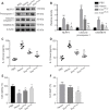
miR-23b negatively regulated PTEN in BV2 cells. (A) Predicted binding between PTEN and miR-23b. (B) Luciferase report assays confirmed binding between miR-23b and PTEN. (C) Relative PTEN mRNA levels after transfection with the mimic and inhibitor. (D) PTEN protein expression after transfection with the mimic and inhibitor. Data are shown as the mean ± SD (n = 3). **P < 0.01 (Student’s t-test). Inhibitors: transfected with miR-23b inhibitors; mimics: transfected with miR-23b mimics; miR-NC: transfected with miR-NC; ns: not significant; PTEN: phosphatase and tensin homolog deleted on chromosome 10.
Figure 10
Figure 10
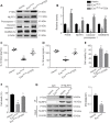
BMSC-exosomal miR-23b attenuates oxidative stress by regulating the PTEN/Nrf2 pathway in microglia BV2 cells in vitro. (A) Western blot bands for PTEN, Nrf2 (cytoplasmic and nuclear), and HO-1 proteins in microglia BV2 cells from different groups. (B) Quantitative analysis of PTEN, Nrf2 (cytoplasmic and nuclear), and HO-1. (C) MDA levels were measured by economical kits. (D) SOD levels were measured by economical kits. (E) GSH/GSSG ratio was calculated on the basis of the GSH and GSSG levels which evaluated by economical kits. Data are shown as the mean ± SD (n = 6). *P < 0.05, **P < 0.01, vs. hemin group; #P < 0.05, vs. exomiR-23b group (one-way analysis of variance followed by Newman-Keuls post hoc analysis). Hemin: Treated with 60 μM hemin for 24 hours; exomiR-23b group: treated with miR-23b transfected BMSCs-exosomes after hemin stimulation; exomiR-23b + PTEN: treated with pcDNA3.1-PTEN plasmids and miR-23b transfected BMSCs-exosomes after hemin stimulation. BMSC: Bone marrow mesenchymal stem cell; GSH: reduced glutathione; GSSG: oxidized glutathione disulfide; HO-1: heme oxygenase-1; MDA: malondialdehyde; Nrf2: nuclear factor erythroid-2-related factor 2; PTEN: phosphatase and tensin homolog deleted on chromosome 10; SOD: superoxide dismutase.
Figure 11
Figure 11
BMSC-exosomal miR-23b alleviates NLRP3 inflammasome-mediated pyroptosis by regulating PTEN in vitro. (A, B) Representative bands and data analysis for PTEN and pyroptosis-related proteins by western blotting in BV2 cells. (C) IL-1β and (D) IL-18 levels in the BV2 cellular supernatant were evaluated using ELISA. (E) Cell viability of hippocampal neuronal HT22 cells was assessed using the CCK8 test. (F) Cell death rates of HT22 cells by LDH assays. (G) Co-IP and data analysis of the interaction between endogenous PTEN and NLRP3 in BV2 cells. Data are shown as the mean ± SD (n = 6).*P < 0.05, **P < 0.01, vs. hemin group; #P < 0.05, ##P < 0.01, vs. exomiR-23b group (one-way analysis of variance with the Newman−Keuls post hoc analysis in A−F; Student’s t-test in G). Hemin: Treated with 60 μM hemin for 24 hours; exomiR-23b group: treated with miR-23b transfected BMSC-exosomes after hemin stimulation; exomiR-23b + PTEN: treated with pcDNA3.1-PTEN plasmids and miR-23b transfected BMSC-exosomes after hemin stimulation. BMSC: Bone marrow mesenchymal stem cell; CCK8: cell counting kit-8; Co-IP: co-immunoprecipitation; ELISA: enzyme linked immunosorbent assay; GSDMD-N: N-terminal fragment of gasdermin D; IL-18: interleukin-18; IL-1β: interleukin-1β; LDH: lactate dehydrogenase; NLRP3: NOD-like receptor family pyrin domain containing 3; PTEN: phosphatase and tensin homolog deleted on chromosome 10.

Similar articles

Cited by

References

    1. Bushati N, Cohen SM. microRNA functions. Annu Rev Cell Dev Biol. 2007;23:175–205. - PubMed
    1. Cacialli P, Mahony CB, Petzold T, Bordignon P, Rougemont AL, Bertrand JY. A connexin/ifi30 pathway bridges HSCs with their niche to dampen oxidative stress. Nat Commun. 2021;12:4484. - PMC - PubMed
    1. Chen W, Guo C, Feng H, Chen Y. Mitochondria:novel mechanisms and therapeutic targets for secondary brain injury after intracerebral hemorrhage. Front Aging Neurosci. 2020a;12:615451. - PMC - PubMed
    1. Chen W, Guo C, Huang S, Jia Z, Wang J, Zhong J, Ge H, Yuan J, Chen T, Liu X, Hu R, Yin Y, Feng H. MitoQ attenuates brain damage by polarizing microglia towards the M2 phenotype through inhibition of the NLRP3 inflammasome after ICH. Pharmacol Res. 2020b;161:105122. - PubMed
    1. Chen X, Zhou Y, Wang S, Wang W. Mechanism of baicalein in brain injury after intracerebral hemorrhage by inhibiting the ROS/NLRP3 inflammasome pathway. Inflammation. 2022;45:590–602. - PubMed