. 2023 Mar;18(3):657-663.
doi: 10.4103/1673-5374.350212.

A hyaluronic acid granular hydrogel nerve guidance conduit promotes regeneration and functional recovery of injured sciatic nerves in rats

Affiliations

A hyaluronic acid granular hydrogel nerve guidance conduit promotes regeneration and functional recovery of injured sciatic nerves in rats

Jie Yang et al. Neural Regen Res. 2023 Mar.

Abstract

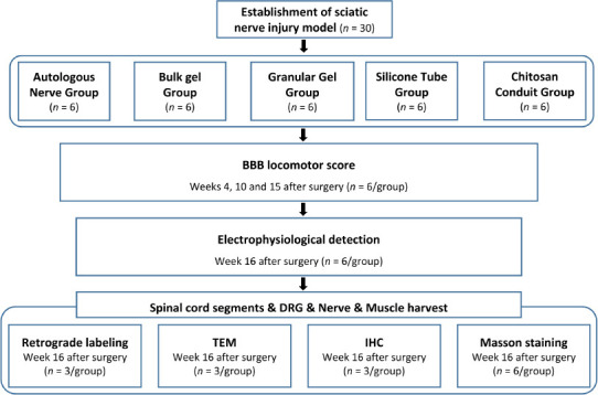
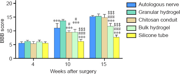
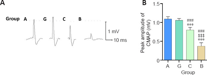
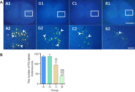
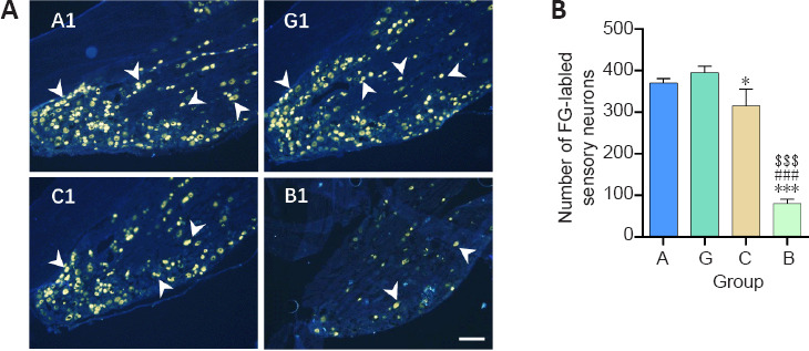
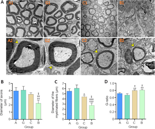
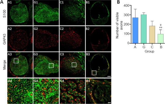
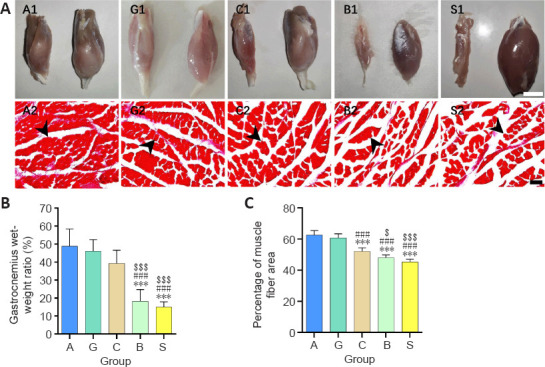
A hyaluronic acid granular hydrogel can promote neuronal and astrocyte colony formation and axonal extension in vitro, suggesting that the hydrogel can simulate an extracellular matrix structure to promote neural regeneration. However, in vivo experiments have not been conducted. In this study, we transplanted a hyaluronic acid granular hydrogel nerve guidance conduit to repair a 10-mm long sciatic nerve gap. The Basso, Beattie, and Bresnahan locomotor rating scale, sciatic nerve compound muscle action potential recording, Fluoro-Gold retrograde tracing, growth related protein 43/S100 immunofluorescence staining, transmission electron microscopy, gastrocnemius muscle dry/wet weight ratio, and Masson's trichrome staining results showed that the nerve guidance conduit exhibited similar regeneration of sciatic nerve axons and myelin sheath, and recovery of the electrophysiological function and motor function as autologous nerve transplantation. The conduit results were superior to those of a bulk hydrogel or silicone tube transplant. These findings suggest that tissue-engineered nerve conduits containing hyaluronic acid granular hydrogels effectively promote the morphological and functional recovery of the injured sciatic nerve. The nerve conduits have the potential as a material for repairing peripheral nerve defects.

Keywords: functional recovery; granular hydrogel; hyaluronic acid; myelin sheath; nerve conduit; nerve regeneration; peripheral nerve regeneration; sciatic nerve injury; tissue engineering; transection injury.


Conflict of interest statement

None

Figures

Figure 1
Figure 1
Flow chart of the experimental procedures and animal use. BBB: Basso, Beattie, and Bresnahan score; DRG: dorsal root ganglion; IHC: immunohistochemistry; TEM: transmission electron microscopy.
Figure 2
Figure 2
Effect of the granular hydrogel nerve guidance conduit on the BBB locomotor scale in rats with sciatic nerve injury. The BBB locomotor scale was applied 4, 10, and 15 weeks after surgery. The higher the score, the better the motor function recovery of the animal. The scores of each group increased over time, and the difference in scores between the groups changed significantly. Data are shown as mean ± SD (n = 6). ***P < 0.001, vs. granular hydrogel group; #P < 0.05, ###P < 0.001, vs. autologous nerve group; $$$P < 0.001, vs. chitosan conduit group (one-way analysis of variance followed by Tukey’s post hoc test). BBB: Basso, Beattie, and Bresnahan score.
Figure 3
Figure 3
Effect of the granular hydrogel nerve guidance conduit on the CMAP of the operation side of the sciatic nerves in rats 16 weeks after surgery. (A) The CMAP waveforms of the autologous nerve, granular hydrogel, chitosan conduit, and bulk gel conduit groups were recorded in the proximal nerve portion of the stimulation. The peak amplitudes of the CMAP waveforms in the autologous nerve and granular hydrogel groups were the greatest, followed by the chitosan conduit group and bulk hydrogel group. (B) Peak amplitude of CMAP. Data are shown as the mean ± SD (n = 6). ***P < 0.001, vs. Group G; ###P < 0.001, vs. Group A; $$$P < 0.001, vs. Group C (one-way analysis of variance followed by Tukey’s post hoc test). CMAP: Compound muscle action potential; Group A: autologous nerve group; Group B: bulk hydrogel group; Group C: chitosan conduit group; Group G: granular hydrogel group.
Figure 4
Figure 4
Effect of the granular hydrogel nerve guidance conduit on the FG-labeled motoneurons in the spinal anterior horn of rats with sciatic nerve injury 16 weeks after surgery. (A) The distribution of the FG-labeled motoneurons (arrowheads) in the spinal anterior horn. Groups G (G1 and G2) and A (A1 and A2) showed more labeled neurons than Groups C (C1 and C2) and B (B1 and B2). A2, G2, C2, and B2 are the zoomed-in fields of A1, G1, C1, and B1, respectively. Scale bars: 1 mm (upper), 200 µm (lower). (B) The number of FG-labeled motoneurons on 10 spinal cord slices from each animal. Data are shown as the mean ± SD (n = 3). **P < 0.01, ***P < 0.001, vs. Group G; ##P < 0.01, ###P < 0.001, vs. Group A; $$$P < 0.001, vs. Group C (one-way analysis of variance followed by Tukey’s post hoc test). FG: Fluoro-gold; Group A: autologous nerve group; Group B: bulk hydrogel group; Group C: chitosan conduit group; Group G: granular hydrogel group.
Figure 5
Figure 5
Effect of the granular hydrogel nerve guidance conduit on the FG-labeled sensory neurons in the DRG of rats with sciatic nerve injury 16 weeks after surgery. (A) The distribution of the FG-labeled sensory neurons (arrowheads) in DRG. Groups G (G1) and A (A1) showed more labeled neurons than Groups C (C1) and B (B1). Scale bar: 200 µm. (B) The number of FG-labeled sensory neurons on three dorsal root ganglion slices from each animal. Data are shown as the mean ± SD (n = 3). *P < 0.05, ***P < 0.001, vs. Group G; ###P < 0.001, vs. Group A; $$$P < 0.001, vs. Group C (one-way analysis of variance followed by Tukey’s post hoc test). FG: Fluoro-gold; Group A: autologous nerve group; Group B: bulk hydrogel group; Group C: chitosan conduit group; Group G: granular hydrogel group.
Figure 6
Figure 6
Effect of the granular hydrogel nerve guidance conduit on the morphology analysis of regenerated nerves in rats with sciatic nerve injury 16 weeks after surgery. (A) The ultrastructure of the regenerated nerves (arrowheads). The diameters of nerve fibers in Groups A (A1 and A2) and G (G1 and G2) were visibly larger and the distribution of myelin sheaths was more compact and uniform than Groups C (C1 and C2) and B (B1 and B2). Scale bars: 5 µm (upper), 1 µm (lower). (B–D) Quantitative results of the diameter of axons (B), the diameter of myelinated fibers (C), and the G-ratio (D) in the distal portion of regenerated nerves. Data are shown as the mean ± SD (n = 3). *P < 0.05, **P < 0.01, ***P < 0.001, vs. Group G; #P < 0.05, ##P < 0.01, ###P < 0.001, vs. Group A; $P < 0.05, vs. Group C (one-way analysis of variance followed by Tukey’s post hoc test). Group A: autologous nerve group; Group B: bulk hydrogel group; Group C: chitosan conduit group; Group G: granular hydrogel group.
Figure 7
Figure 7
Effect of the granular hydrogel nerve guidance conduit on the regenerated nerves in rats with sciatic nerve injury 16 weeks after the surgery. (A) The expression of S100 (green, Alexa Fluor 488) and GAP43 (red, Alexa Fluor 594) in the transverse sections of regenerated nerves. Visibly more myelin sheaths and axons were regenerated in Groups G (G1–G4) and A (A1–A4), followed by Groups C (C1–C4) and B (B1–B4). Scale bars: 100 µm (upper three rows), 20 µm (lowest row). A4, G4, C4, and B4 are the zoomed-in fields of A3, G3, C3, and B3, respectively. (B) Quantification of the number of visible axons. Data are shown as the mean ± SD (n = 3). **P < 0.01, vs. Group G; #P < 0.05, vs. Group A (one-way analysis of variance followed by Tukey’s post hoc test). GAP43: Growth associated protein 43; Group A: autologous nerve group; Group B: bulk hydrogel group; Group C: chitosan conduit group; Group G: granular hydrogel group.
Figure 8
Figure 8
Effect of the granular hydrogel nerve guidance conduit on the gastrocnemius muscle atrophy in rats with sciatic nerve injury 16 weeks after surgery. (A) Gross appearance of the gastrocnemius muscles on the injured (left) and normal (right) sides (A1, G1, C1, B1, and S1). Masson’s staining of gastrocnemius muscles in each group (A2, G2, C2, B2, and S2). The gastrocnemius muscle of the injured side was visibly smaller than that of the normal side and Groups A and G showed more muscle fibers and less spacing between muscle fibers in the same view size field. Arrowheads indicate the positive muscle fibers. Scale bars: 1 cm (upper) and 100 µm (lower). (B and C) Comparisons of the gastrocnemius wet weight ratio (B) and the average percentage of the muscle fiber area (C). Data are shown as the mean ± SD (n = 6). ***P < 0.001, vs. Group G; ###P < 0.001, vs. Group A; $P < 0.05, $$$P < 0.001, vs. Group C (one-way analysis of variance followed by Tukey’s post hoc test). Group A: autologous nerve group; Group B: bulk hydrogel group; Group C: chitosan conduit group; Group G: granular hydrogel group.

References

    1. Basso DM, Beattie MS, Bresnahan JC. A sensitive and reliable locomotor rating scale for open field testing in rats. J Neurotrauma. 1995;12:1–21. - PubMed
    1. Biazar E. Design of porous alginate conduit using a freeze drying technique for neural engineering. Orient J Chem. 2011;27:1029–1032.
    1. Carvalho CR, Oliveira JM, Reis RL. Modern trends for peripheral nerve repair and regeneration:beyond the hollow nerve guidance conduit. Front Bioeng Biotechnol. 2019;7:337. - PMC - PubMed
    1. Chen Y, Zhao C, Zhang C, Luo L, Yu G. Influence of chronic intermittent hypoxia on growth associated protein 43 expression in the hippocampus of young rats. Neural Regen Res. 2012;7:1241–1246. - PMC - PubMed
    1. Chen ZL, Strickland S. Laminin gamma1 is critical for Schwann cell differentiation, axon myelination, and regeneration in the peripheral nerve. J Cell Biol. 2003;163:889–899. - PMC - PubMed