

Congenital sacrococcygeal rhabdomyosarcoma
- PMID: 36018208
- PMCID: PMC9615950
- DOI: 10.4103/ajps.ajps_69_21
Congenital sacrococcygeal rhabdomyosarcoma
Abstract
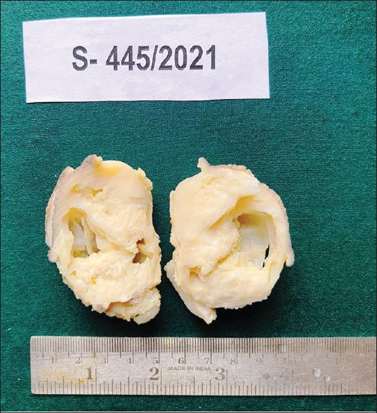
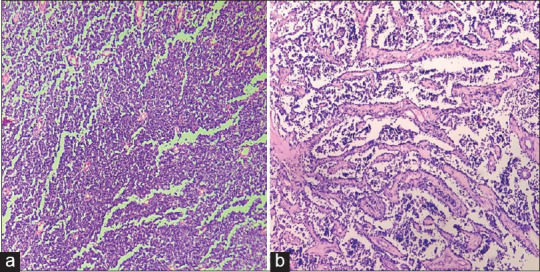
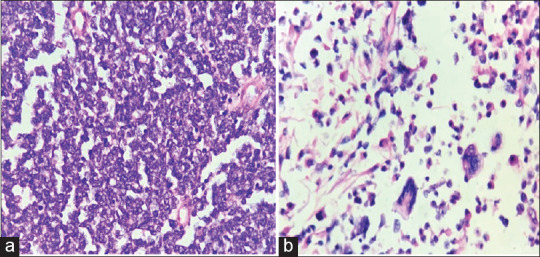
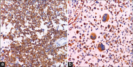
Rhabdomyosarcoma (RMS) is one of the common malignant soft-tissue sarcomas affecting children. It originates from the embryonic mesenchyme precursor of striated muscle and is frequently seen in the head-and-neck region, genitourinary system and extremities. Occasionally, it arises from the retroperitoneum, biliary tract and abdomen and is rarely seen in the sacrococcygeal area. A 4-month-male child presented with a nodule over the sacrum. Based on histopathology and immunohistochemical marker studies, a final diagnosis of RMS was rendered. There was no evidence of any teratomatous elements.
Keywords: Congenital; desmin; immunohistochemistry; infants; rhabdomyosarcoma; sacrococcygeal tumour.
Conflict of interest statement
None
Figures
References
-
- Burrows NP, Ratnavel RC, Grant JW, Cormack GC, Pye RJ. Auricular embryonal rhabdomyosarcoma. Dermatology. 2014;189:301–3. - PubMed
-
- Chirat M, Dainese L, Fasola S, Couloigner V, Denoyelle F, Garabedian EN, et al. Unusual outer ear swelling: Childhood auricular rhabdomyosarcoma. Eur Ann Otorhinolaryngol Head Neck Dis. 2016;133:23–6. - PubMed
-
- Brecher AR, Reyes-Mugica M, Kamino H, Chang MW. Congenital primary cutaneous rhabdomyosarcoma in a neonate. Pediatr Dermatol. 2003;20:335–8. - PubMed
Publication types
MeSH terms
LinkOut - more resources
Full Text Sources
