

Isolated duodenal duplication cyst in a neonate
- PMID: 36018210
- PMCID: PMC9615949
- DOI: 10.4103/ajps.ajps_176_21
Isolated duodenal duplication cyst in a neonate
Abstract
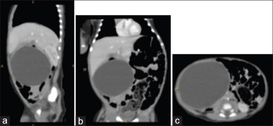
Duodenal duplication cysts are a rare subtype of alimentary tract duplications cysts, consisting of 7% of all the duplications. We report a rare case of neonatal duodenal duplication cyst presenting as a palpable abdominal mass and features of gastric outlet obstruction. A 27-day-old male child presented with complaints of icterus, non-bilious vomiting after every feed and right-sided abdominal lump for the last 15 days. A computed tomography scan of the abdomen revealed well-defined peripherally enhancing cystic lesion noted in the subhepatic region extending up to the right lumbar region. On surgical exploration, a cystic mass was found attached to the pyloric part of the stomach along the mesenteric border of the first, second and third part of the duodenum, which was marsupialised, and no communication was found with the duodenum. On histopathological analysis, a duodenal duplication cyst was diagnosed without any heterotopic mucosa. The literature was reviewed and the approach to duodenal duplication cyst in neonates is discussed.
Keywords: Duodenum; duodenal duplication cyst; neonatal abdominal lump; neonate; paediatric surgery.
Conflict of interest statement
None
Figures


Similar articles
-
Diagnosis and surgical management of a rare case of duodenal duplication cyst in a neonate: Case report and literature review.Int J Surg Case Rep. 2023 Jun;107:108354. doi: 10.1016/j.ijscr.2023.108354. Epub 2023 May 30. Int J Surg Case Rep. 2023. PMID: 37267789 Free PMC article.
-
Isolated duodenal duplication cyst presenting as a complex solid and cystic mass in the upper abdomen.J Radiol Case Rep. 2013 Nov 1;7(11):32-7. doi: 10.3941/jrcr.v7i11.1785. eCollection 2013 Nov. J Radiol Case Rep. 2013. PMID: 24421928 Free PMC article. Review.
-
Duodenal duplication cyst: a potentially malignant disease.Ann Surg Oncol. 2012 Nov;19(12):3753-4. doi: 10.1245/s10434-012-2502-4. Epub 2012 Jul 26. Ann Surg Oncol. 2012. PMID: 22832999
-
Sonographic findings in a duodenal duplication cyst.J Clin Ultrasound. 2002 Nov-Dec;30(9):566-8. doi: 10.1002/jcu.10117. J Clin Ultrasound. 2002. PMID: 12404525
-
Meta-analysis: the clinical features of the duodenal duplication cyst.J Pediatr Surg. 2010 Aug;45(8):1598-606. doi: 10.1016/j.jpedsurg.2010.01.010. J Pediatr Surg. 2010. PMID: 20713206 Review.
Cited by
-
Duodenal duplication cyst causing partial obstruction in a neonate.JPGN Rep. 2025 May 13;6(3):322-324. doi: 10.1002/jpr3.70028. eCollection 2025 Aug. JPGN Rep. 2025. PMID: 40814590 Free PMC article. No abstract available.
References
-
- Mba, M. I. G. H. W., Md, P. M. J., & Peter, S. S. D., MD. (2019). Ashcraft's Pediatric Surgery: Expert Consult-Online+Print (7th ed.) [E-book]. Elsevier
-
- Macpherson RI. Gastrointestinal tract duplications: Clinical, pathologic, etiologic, and radiologic considerations. Radiographics. 1993;13:1063–80. - PubMed
-
- Edwards H. Congenital diverticula of the intestine: With report of a case exhibiting heterotopia. Br J Surg. 1929;17:7–21.
-
- Lewis FT, Thyng FW. The regular occurrence of intestinal diverticula in embryos of the pig, rabbit, and man. Am J Anat. 1908;7:505–519.
Publication types
MeSH terms
LinkOut - more resources
Full Text Sources
Medical
Molecular Biology Databases
