

Potential mechanisms for lung fibrosis associated with COVID-19 infection
- PMID: 36018274
- PMCID: PMC10382189
- DOI: 10.1093/qjmed/hcac206
Potential mechanisms for lung fibrosis associated with COVID-19 infection
Abstract
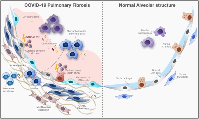
Pulmonary fibrosis is a sequelae of Severe Acute Respiratory Syndrome Coronavirus 2 (SARS-CoV-2) infection that currently lacks effective preventative or therapeutic measures. Post-viral lung fibrosis due to SARS-CoV-2 has been shown to be progressive on selected patients using imaging studies. Persistent infiltration of macrophages and monocytes, a main feature of SARS-CoV-2 pulmonary fibrosis, and long-lived circulating inflammatory monocytes might be driving factors promoting the profibrotic milieu in the lung. The upstream signal(s) that regulates the presence of these immune cells (despite complete viral clearance) remains to be explored. Current data indicate that much of the stimulating signals are localized in the lungs. However, an ongoing low-grade systemic inflammation in long Coronavirus Disease 2019 (COVID-19) symptoms suggests that certain non-pulmonary regulators such as epigenetic changes in hematopoietic stem cells might be critical to the chronic inflammatory response. Since nearly one-third of the world population have been infected, a timely understanding of the underlying pathogenesis leading to tissue remodeling is required. Herein, we review the potential pathogenic mechanisms driving lung fibrosis following SARS-CoV-2 infection based upon available studies and our preliminary findings (Graphical abstract).
© The Author(s) 2022. Published by Oxford University Press on behalf of the Association of Physicians. All rights reserved. For permissions, please email: journals.permissions@oup.com.
Figures
Similar articles
-
Post-COVID pulmonary sequelae: Mechanisms and potential targets to reduce persistent fibrosis.Pharmacol Ther. 2025 Aug;272:108891. doi: 10.1016/j.pharmthera.2025.108891. Epub 2025 May 28. Pharmacol Ther. 2025. PMID: 40447142 Review.
-
Pathogenesis-directed therapy of 2019 novel coronavirus disease.J Med Virol. 2021 Mar;93(3):1320-1342. doi: 10.1002/jmv.26610. Epub 2020 Nov 10. J Med Virol. 2021. PMID: 33073355 Review.
-
A phosphodiesterase-4 inhibitor reduces lung inflammation and fibrosis in a hamster model of SARS-CoV-2 infection.Front Immunol. 2023 Oct 2;14:1270414. doi: 10.3389/fimmu.2023.1270414. eCollection 2023. Front Immunol. 2023. PMID: 37854602 Free PMC article.
-
Pulmonary fibrosis in COVID-19: mechanisms, consequences and targets.QJM. 2023 Oct 6;116(9):750-754. doi: 10.1093/qjmed/hcad092. QJM. 2023. PMID: 37191984 Review.
-
Cytokines and Lipid Mediators of Inflammation in Lungs of SARS-CoV-2 Infected Mice.Front Immunol. 2022 Jun 24;13:893792. doi: 10.3389/fimmu.2022.893792. eCollection 2022. Front Immunol. 2022. PMID: 35812400 Free PMC article.
Cited by
-
Unveiling the Pathological Mechanisms of Death Induced by SARS-CoV-2 Viral Pneumonia.Microorganisms. 2024 Feb 24;12(3):459. doi: 10.3390/microorganisms12030459. Microorganisms. 2024. PMID: 38543510 Free PMC article.
-
Targeted immunotherapy rescues pulmonary fibrosis by reducing activated fibroblasts and regulating alveolar cell profile.Nat Commun. 2025 Apr 21;16(1):3748. doi: 10.1038/s41467-025-59093-7. Nat Commun. 2025. PMID: 40258811 Free PMC article.
-
Maladaptive TGF-β Signals to the Alveolar Epithelium Drive Fibrosis after COVID-19 Infection.Am J Respir Crit Care Med. 2023 Jul 15;208(2):201-204. doi: 10.1164/rccm.202302-0264LE. Am J Respir Crit Care Med. 2023. PMID: 37236627 Free PMC article. No abstract available.
-
Wogonin protects against bleomycin-induced mouse pulmonary fibrosis via the inhibition of CDK9/p53-mediated cell senescence.Front Pharmacol. 2024 Jul 8;15:1407891. doi: 10.3389/fphar.2024.1407891. eCollection 2024. Front Pharmacol. 2024. PMID: 39040475 Free PMC article.
-
Morphopathology of the lesions induced by SARS-CoV-2 infection in the lungs.Rom J Morphol Embryol. 2024 Oct-Dec;65(4):637-645. doi: 10.47162/RJME.65.4.10. Rom J Morphol Embryol. 2024. PMID: 39957025 Free PMC article.
References
Publication types
MeSH terms
Grants and funding
LinkOut - more resources
Full Text Sources
Medical
Miscellaneous
